

Thyroarytenoid
By
Della Barnes, an MS Anatomy graduate, blends medical research with accessible writing, simplifying complex anatomy for a better understanding and appreciation of human anatomy.
Last updated:
09/01/2026Della Barnes, MS Anatomy
UX/UI Designer at - AdobeDella Barnes, an MS Anatomy graduate, blends medical research with accessible writing, simplifying complex anatomy for a better understanding and appreciation of human anatomy.
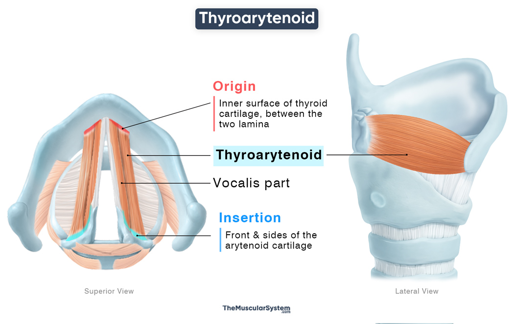
The thyroarytenoid is a narrow, paired muscle on the inner side of the larynx. It is one of the intrinsic laryngeal muscles, helping with vocalization along with the other intrinsic muscles of the larynx, the cricothyroid, posterior cricoarytenoid, lateral cricoarytenoid, oblique arytenoid, and transverse arytenoid. It is mainly involved in managing the tension on the vocal cords to control pitch during vocalization.
Anatomy
Location and Attachments
| Origin | Inner surface of the thyroid cartilage, between the two lamina |
| Insertion | Front and sides of the arytenoid cartilage |
Origin
The muscle originates from the inner surface of the thyroid cartilage, just from the angle formed by the joining of the two lamina. Part of the muscle also rises from the cricothyroid ligament, the connection between the thyroid cartilage and the cricoid cartilage.
Insertion
From the point of origin, the muscle fibers course in multiple directions, mainly along the lateral sides of the larynx. The majority of the fibers course slightly obliquely towards the front as they form the muscle belly. This part inserts into the anterior and lateral sides of the arytenoid cartilage.
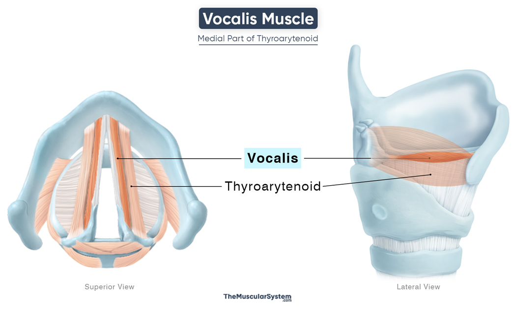
The inner, deeper fibers run parallel with the vocal ligament and insert into the front of the arytenoid cartilage, specifically into the vocal processes. This portion is sometimes recognized separately as the vocalis muscle.
One portion of the muscle fibers extends to blend with the aryepiglottic fold, while some reach the borders of the epiglottis. This part may also be considered as a small yet separate muscle, named the thyroepiglottic or thyreoepiglotticus muscle.
Relations With Surrounding Muscles and Structures
Despite its small size, the thyroarytenoid is an important structure. Its medial fibers, or the vocalis part, constitute the muscular layer of the vocal folds.
The muscle contributes to the medial wall of the laryngeal ventricle, a fusiform recess located between the vestibular and vocal folds and lined by respiratory epithelium containing mucous glands. Superior to the ventricle lies the laryngeal saccule, a mucosal extension that lubricates the vocal folds.
Function
| Action | Relaxing the vocal cords to help with vocalization and pitch control |
It is the primary relaxer of the vocal cords. Working together with the rest of the laryngeal muscles, mainly the primary tensor of the vocal cords, the cricothyroid, the thyroarytenoid plays a vital role in vocalization
Effect on pitch
When the muscle contracts, it adducts the vocal ligament and pulls the arytenoid cartilage anteriorly toward the thyroid cartilage, shortening and relaxing the vocal folds. This reduces their longitudinal tension, causing them to vibrate more slowly, producing lower-pitched sounds.
Fine control of phonation (vocalis part)
Through contraction of its medial fibers (the vocalis part), the muscle adjusts tension in specific segments of the vocal fold, allowing fine control of pitch during phonation.
Role in glottic closure
Due to its fiber orientation, the muscle is also able to assist the laryngeal adductors like lateral cricoarytenoid, helping in closing the anterior part of the rima glottidis by medially rotating the arytenoid cartilage and adducting the vocal folds.
Antagonists
The cricothyroid muscle acts as its primary antagonist by increasing tension on the vocal folds, thereby counteracting the thyroarytenoid’s role in relaxing them.
Innervation
| Nerve | Recurrent laryngeal nerve |
The muscle, along with its vocalis part, is innervated by the recurrent laryngeal nerve, which branches off of the vagus nerve (CN X). Additional nerve supply comes from the external laryngeal branch of the superior laryngeal nerve, another branch of the vagus nerve.
Blood Supply
| Artery | Superior and inferior thyroid arteries |
The laryngeal branches of the superior and inferior thyroid arteries provide blood supply to this muscle. The two arteries originate from the external carotid artery and the thyrocervical trunk, respectively.
References
- Thyroarytenoid Muscle: Kenhub.com
- Thyroarytenoid Muscle: Elsevier.com
- Thyroarytenoid: TeachMeAnatomy.info
- Vocalis Muscle: IMIAIOS.com
- Intrinsic Muscles of the Larynx: Radiopaedia.org
Della Barnes, an MS Anatomy graduate, blends medical research with accessible writing, simplifying complex anatomy for a better understanding and appreciation of human anatomy.
- Latest Posts by Della Barnes, MS Anatomy
-
Tensor Tympani
- -
Stapedius
- -
Auricularis Posterior
- All Posts


