

Stylopharyngeus
By
Della Barnes, an MS Anatomy graduate, blends medical research with accessible writing, simplifying complex anatomy for a better understanding and appreciation of human anatomy.
Last updated:
22/12/2025Della Barnes, MS Anatomy
UX/UI Designer at - AdobeDella Barnes, an MS Anatomy graduate, blends medical research with accessible writing, simplifying complex anatomy for a better understanding and appreciation of human anatomy.
What is the Stylopharyngeus
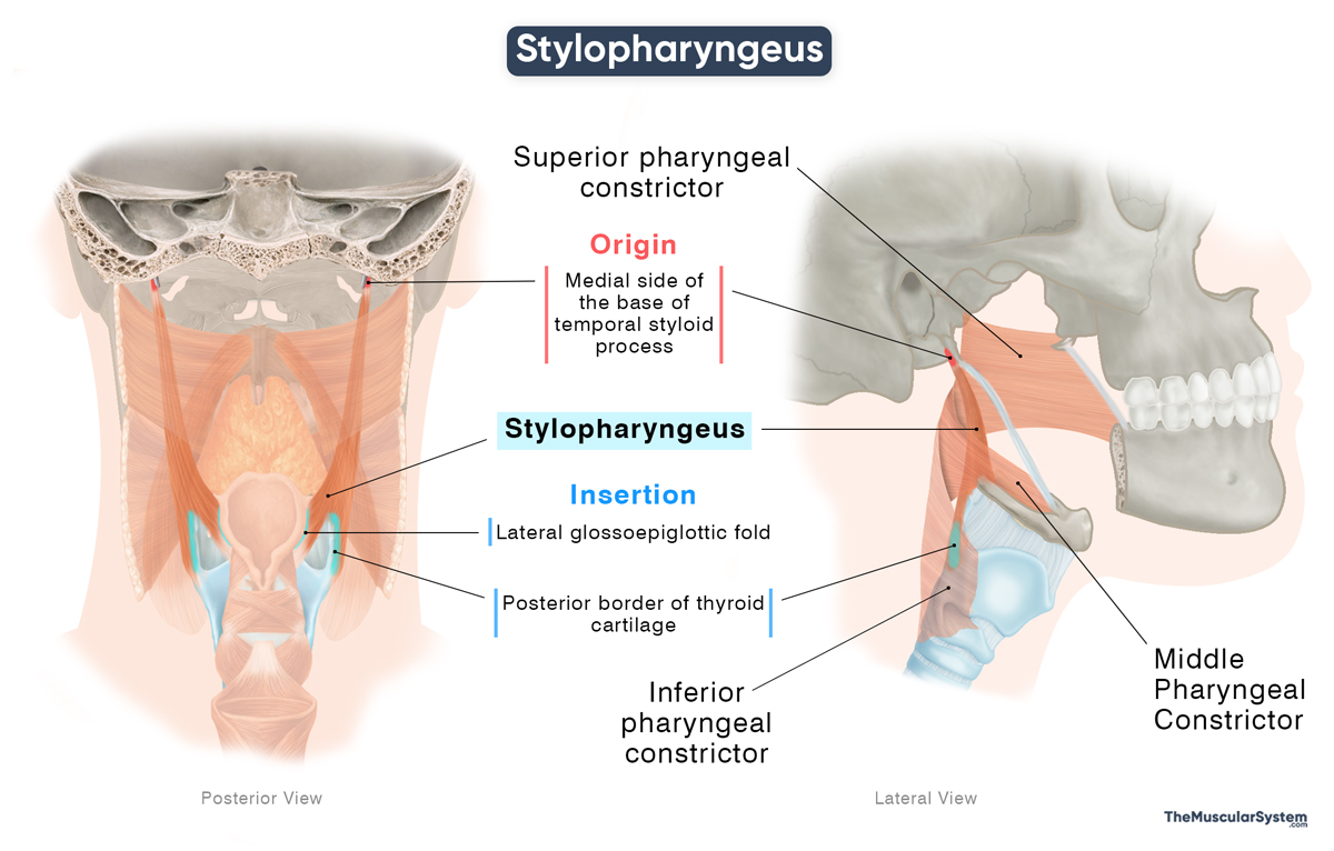
The stylopharyngeus is a thin, long, paired muscle in the neck, located on either side of the pharynx. It belongs to the group of longitudinal pharyngeal muscles, along with the salpingopharyngeus and palatopharyngeus muscles. The muscle plays a vital role in swallowing by helping elevate the pharynx and larynx.
Anatomy
Location and Attachments
| Origin | Medial base of the temporal styloid process |
| Insertion | Posterior margin of the thyroid cartilage, lateral glossoepiglottic fold; some fibers blend with the superior and middle pharyngeal constrictors |
Origin
The muscle originates from the medial aspect of the base of the styloid process of the temporal bone. This makes it the only pharyngeal muscle that does not attach to the pharyngeal wall.
Insertion
After originating, the muscle fibers course inferiorly, forming a cylindrical muscle belly. As the muscle approaches the pharyngeal mucosa, its fibers spread out, becoming flat, like a band. It then inserts into three points:
- Some of the fibers blend with the superior and middle pharyngeal constrictors.
- Part of the muscle inserts into the lateral glossoepiglottic fold, a paired mucosal fold in the throat, on the sides of the epiglottis
- The remaining fibers blend with the inserting fibers of the palatopharyngeus muscle and attach to the posterior margin of the thyroid cartilage.
Relations With Surrounding Muscles and Structures
As it descends from its point of origin, it passes through the gap between the superior and middle pharyngeal constrictor muscles, along with the glossopharyngeal nerve (CN IX) and the stylohyoid ligament. The glossopharyngeal nerve curves around the posterior border of the muscle and then runs along its lateral surface toward the tongue. Deep to the pharyngeal constrictors, the muscle is invested by the pharyngobasilar fascia.
The ascending pharyngeal artery and the palatopharyngeus muscle run posterior and lateral to this muscle. Above the digastric muscle, the stylopharyngeus courses between the external carotid artery from the internal carotid artery. Near the parotid gland, it lies between the external carotid artery and deeper structures, including the internal jugular vein and vagus nerve (CN X).
At the level of the tensor veli palatini, the stylopharyngeus lies between the medial pterygoid muscle and the superior pharyngeal constrictor, contributing to the lateral wall of the pharynx.
Functionally, together with the palatopharyngeus, the stylopharyngeus contributes to the lateral wall of the piriform recess, an important pathway for swallowing and vocalization.
Function
| Action | Elevating the pharynx and larynx to help with swallowing |
During swallowing, when food reaches the pharynx, the pharyngeal constrictor muscles push the food downward. After this, the stylopharyngeus, along with other longitudinal muscles, lifts and shortens the pharynx. This makes it easier for the food to move from the pharynx into the esophagus and helps clear food from the throat.
During inhalation, it pulls the pharyngeal wall slightly outward. This helps keep the airway open and prevents the soft pharyngeal wall from collapsing while air is being drawn into the lungs.
During speech, this muscle helps change the shape of the vocal tract by lifting the pharynx and larynx, helping control voice resonance.
Antagonists
The stylopharyngeus has no direct antagonist. Its action ends when it relaxes, after which the pharynx returns to its resting position as the infrahyoid muscles depress the larynx, indirectly lowering the pharynx.
Innervation
| Nerve | Glossopharyngeal nerve (CN IX) |
It is the only pharyngeal muscle innervated by the glossopharyngeal nerve (CN IX); all others are innervated by the vagus nerve (CN X).
Blood Supply
| Artery | Pharyngeal trunk of the ascending pharyngeal artery |
Blood supply to this muscle comes from the pharyngeal trunk of the ascending pharyngeal artery, which itself is a branch of the external carotid artery.
References
- Anatomy, Head and Neck, Stylopharyngeus Muscles: NCBI.NLM.NIH.gov
- Stylopharyngeus: TeachMeAnatomy.info
- Stylopharyngeus: Kenhub.com
- Stylopharyngeus: Meddean.LUC.edu
- Stylopharyngeus Muscle: Radiopaedia.org
- Stylopharyngeus Muscle: Elsevier.com
Della Barnes, an MS Anatomy graduate, blends medical research with accessible writing, simplifying complex anatomy for a better understanding and appreciation of human anatomy.
- Latest Posts by Della Barnes, MS Anatomy
-
Tensor Tympani
- -
Stapedius
- -
Auricularis Posterior
- All Posts


