

Obliquus Capitis Inferior
By
Della Barnes, an MS Anatomy graduate, blends medical research with accessible writing, simplifying complex anatomy for a better understanding and appreciation of human anatomy.
Last updated:
25/11/2025Della Barnes, MS Anatomy
UX/UI Designer at - AdobeDella Barnes, an MS Anatomy graduate, blends medical research with accessible writing, simplifying complex anatomy for a better understanding and appreciation of human anatomy.
What is the Obliquus Capitis Inferior
The obliquus capitis inferior is a paired muscle located at the back of the neck and the base of the skull. It is the largest of the four suboccipital muscles, with the others being the obliquus capitis superior, rectus capitis posterior major, and rectus capitis posterior minor. The muscle helps stabilize and move the head and neck.
Anatomy
Location and Attachments
| Origin | Spinous process of the axis bone |
| Insertion | Transverse process of the atlas bone |
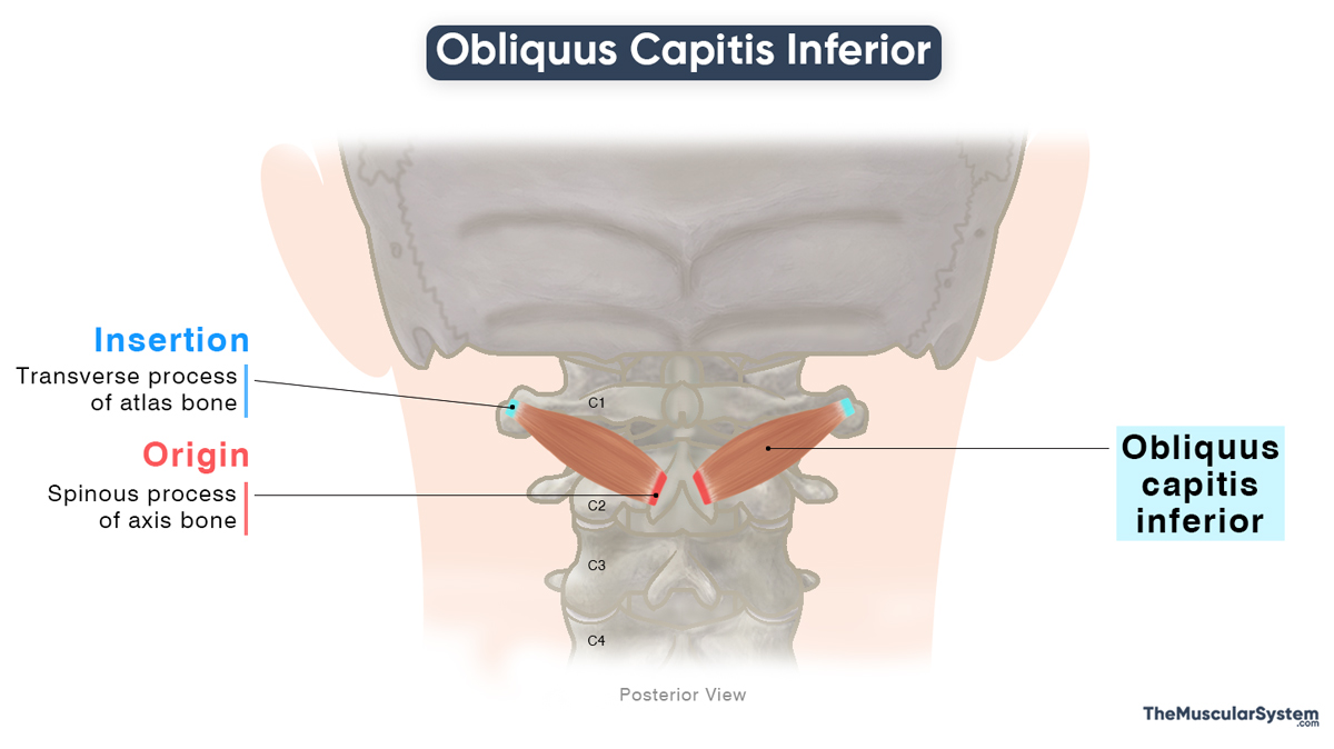
Origin
The muscle originates via a narrow tendon from the inner surface of the spinous process of the second cervical (C2) vertebra, called the axis.
Insertion
After originating, the muscle fibers course laterally upward, forming a short yet broad muscle belly. Although its name, obliquus capitis, suggests a connection to the head — since “capitis” means “of the head” — this muscle does not attach to the skull. Instead, it extends to the atlas, the first cervical vertebra, and inserts on the posterior aspect of its transverse process.
Relations With Surrounding Muscles and Structures
The largest of the suboccipital muscles, the obliquus capitis inferior, lies just below the obliquus capitis superior, which is where its name comes from. It forms the lower lateral boundary of the occipital triangle, an anatomical space at the base of the skull that allows passage for neurovascular structures such as the vertebral artery and the suboccipital nerve.
The muscle also lies deep to the intrinsic back muscles, including the semispinalis capitis and splenius capitis, as well as beneath the upper portion of the trapezius, a superficial back muscle.
Function
| Action | Extending, ipsilaterally rotating, and stabilizing the head and neck at the atlanto-axial joint |
Role in Neck Movement
- When the muscle contracts bilaterally, or on both sides, it extends the atlanto-axial joint, helping lift the head, such as when looking upward.
- When the muscle contracts unilaterally, or on one side, it works with the rectus capitis posterior major to rotate the head ipsilaterally, or toward the same side as the contracting muscle.
The oblique orientation of its muscle fibers, combined with the length of the transverse process of the atlas where it inserts, makes it a powerful neck rotator.
Role in Neck Stability
Like the other suboccipital muscles, this muscle also plays an important role in stabilizing the atlanto-axial joint, helping to keep the head stable on the neck during movement and at rest.
Innervation
| Nerve | Suboccipital nerve |
Innervation to this muscle comes from the suboccipital nerve, which rises from the dorsal ramus of the first cervical spinal nerve (C1).
Blood Supply
| Artery | Vertebral artery and deep descending branch of the occipital artery |
The obliquus capitis inferior receives its blood supply primarily from the vertebral artery, a branch of the subclavian artery, and from the deep portion of the descending branch of the occipital artery, which arises from the external carotid artery.
References
- Obliquus Capitis Inferior Muscle: Kenhub.com
- Obliquus Capitis Inferior: TeachMeAnatomy.info
- Obliquus Capitis Inferior Muscle: Sciencedirect.com
- Obliquus Inferior Capitis Muscle: Elsevier.com
- Obliquus Capitis Inferior Muscle: GetBodySmart.com
Della Barnes, an MS Anatomy graduate, blends medical research with accessible writing, simplifying complex anatomy for a better understanding and appreciation of human anatomy.
- Latest Posts by Della Barnes, MS Anatomy
-
Tensor Tympani
- -
Stapedius
- -
Auricularis Posterior
- All Posts


