Rotatores
Last updated:
30/10/2024Della Barnes, an MS Anatomy graduate, blends medical research with accessible writing, simplifying complex anatomy for a better understanding and appreciation of human anatomy.
What are the Rotatores
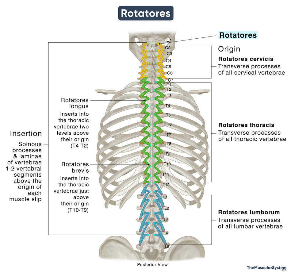
The rotatores are a group of small muscles located along both sides of the vertebral column, playing an important role in stabilizing the spine, as well as assisting with its extension and rotation. They are the smallest muscles in the transversospinalis group and form the deep layer of the intrinsic or deep back muscles, alongside the other muscles in the group transversospinalis, the semispinalis, and multifidus.
Anatomy
Location and Attachments
Like the other transversospinales muscles, the rotatores can also be divided into the cervical, thoracic, and lumbar parts based on the origin of the fibers.
| Origin | Transverse processes of all cervical, thoracic, and lumbar vertebrae |
| Insertion | Spinous process and laminae of vertebrae 1-2 vertebral segments above the origin of each muscular strip or fascicle |
Origin
The muscle fibers from the transverse processes of the cervical vertebrae are called the rotatores cervicis or rotatores colli. Those from the thoracic vertebrae are known as the rotatores thoracis, and the fibers that arise from the lumbar vertebrae are referred to as the rotatores lumborum. However, since the thoracic part is the most prominent, anatomical descriptions often focus primarily on this region.
The fibers of the rotatores thoracis can be divided into two groups: rotatores longus (long rotators) and rotatores brevis (short rotators). Both originate from the superior and dorsal aspects of the transverse processes of the thoracic vertebrae.
Insertion
The muscle fibers run superomedially in an oblique direction, inserting into the spinous processes of vertebrae situated 1-2 levels above the origin of each fascicle.
In the thoracic region, the long rotatores extend two vertebral levels upward to insert into the laminae and spinous processes of thoracic vertebrae located two levels above their point of origin. In contrast, the short rotatores travel a shorter distance, inserting into the laminae and spinous processes of the vertebra immediately above. So, as their names imply, the rotatores longus are longer and more prominent than the rotatores brevis.
Anatomical Variation
Although theoretically, the rotatores muscles are expected to arise from the transverse processes of each vertebra, this is not always the case. In the cervical and lumbar regions, the fascicles can be quite irregular. Anatomical variations exist, where, in some areas, the rotatores are replaced by deeper muscle fibers of the multifidus.
Relations With Surrounding Muscles and Structures
The rotatores muscles make up the deepest layer of the transversospinales group, with the multifidus muscles lying immediately superior to it. Deep to it lie the deepest muscles in the back, the interspinales, intertransversarii, and levatores costarum.
On the spine and thoracic cage, the rotatores are medial to the external intercostals and levatores costarum. Additionally, the entire erector spinae group lies lateral to the rotatores as well.
Function
| Action | Stabilizing the vertebral column, extending and rotating the thoracic spine |
Since the cervicis and lumborum regions are irregular or even absent in some individuals, they have limited impact on the muscle’s functions. The muscle slips in the thoracic part play a more important role. When they contract bilaterally, they help extend the thoracic spine. Conversely, when only one side contracts, it rotates the thoracic spine to the opposite side of the contracting muscle (contralateral contraction).
However, the muscle’s position on the spine limits its mechanical leverage to move the spinal column. In fact, medical literature identifies the primary function of the muscle as stabilizing the spine and maintaining flexibility by supporting the joints between the vertebrae.
Innervation
| Nerve | Dorsal rami of corresponding spinal nerves |
The rotatores muscle receives innervation from the medial branches of the dorsal (or posterior) rami of the spinal nerves corresponding to its level. Therefore, the cervical, thoracic, and lumbar regions of the muscle are innervated by the cervical, thoracic, and lumbar spinal nerves, respectively.
Blood Supply
| Artery | Posterior branches of the posterior intercostal and lumbar arteries |
The posterior branches of the posterior intercostal and lumbar arteries supply these muscles. These arteries arise from the supreme intercostal artery and the aorta, respectively.
References
- Rotatores: TeachMeAnatomy.info
- Rotatores Muscles | Origin, Insertion & Action: Study.com
- Rotatores (Left): Elsevier.com
- Rotatores Muscles: Kenhub.com
- Rotatores Thoracis: IMAIOS.com
Della Barnes, an MS Anatomy graduate, blends medical research with accessible writing, simplifying complex anatomy for a better understanding and appreciation of human anatomy.
- Latest Posts by Della Barnes, MS Anatomy
-
Tensor Tympani
- -
Stapedius
- -
Auricularis Posterior
- All Posts


