

Interspinales
By
Della Barnes, an MS Anatomy graduate, blends medical research with accessible writing, simplifying complex anatomy for a better understanding and appreciation of human anatomy.
Last updated:
04/11/2024Della Barnes, MS Anatomy
UX/UI Designer at - AdobeDella Barnes, an MS Anatomy graduate, blends medical research with accessible writing, simplifying complex anatomy for a better understanding and appreciation of human anatomy.
What Are the Interspinales
The interspinales are small paired muscles in the back, belonging to the deepest layer of intrinsic back muscles. They span between the spinous processes of adjacent vertebrae, which gives them their name “interspinales.” Their primary function is to help maintain the stability and flexibility of the spine.
Anatomy
Location and Attachments
| Origin | Superior surface of the spinous processes starting from C3 to L5 vertebrae |
| Insertion | Inferior surface of the spinous processes starting from C2 to L4 vertebrae |
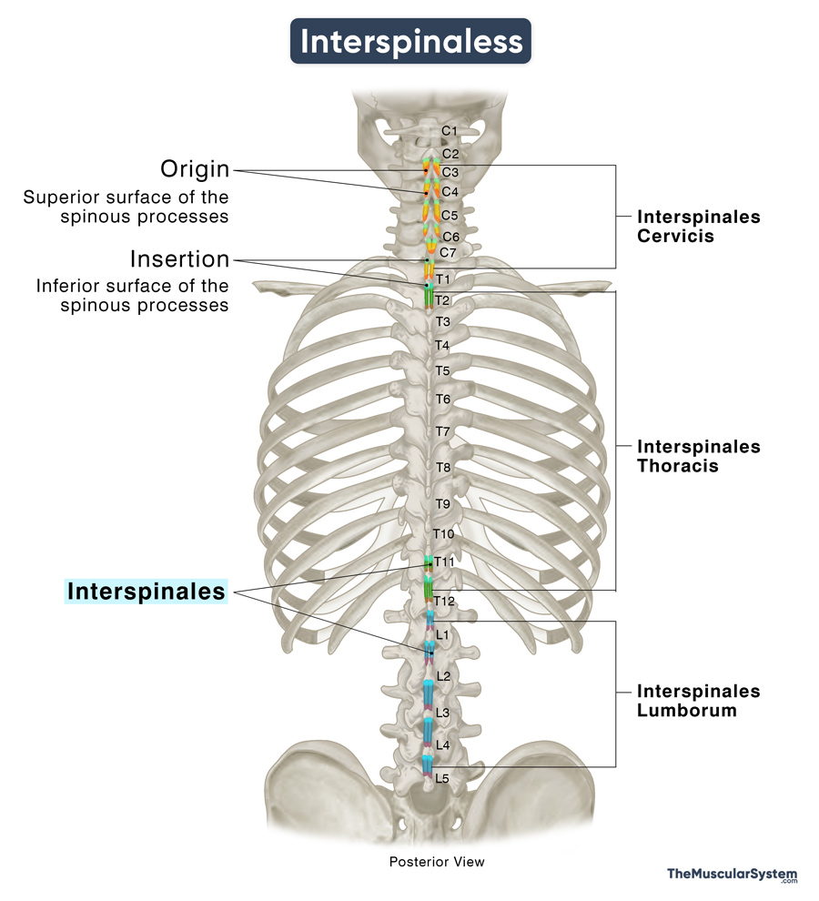
Small muscular fascicles span the upper and lower surfaces of adjacent spinous processes from the cervical to lumbar vertebrae. These fascicles are typically paired, with one located on each side of the interspinous ligament — a small ligament running between the spinous processes along the vertebral column.
Like most other intrinsic back muscles, they can be divided into cervical, thoracic, and lumbar regions based on their origin.
Interspinales Cervicis
It is the most developed part of the interspinales muscle, consisting of 6 distinct pairs originating from the upper surface of the spinous process of the 3rd cervical to 1st thoracic vertebrae (C3-T1). Each fascicle then courses superiorly to insert into the lower surface of the 2nd to the 7th cervical vertebrae (C2 or axis to C7), respectively.
So, the 1st pair of interspinales cervicis is located between the C3 and axis; the 2nd pair is between the C4 and C3, and so on through to C7 and T1.
Interspinales Thoracis
The thoracic region of the muscle is generally the least developed, with fascicles often missing in most thoracic vertebrae. Typically, there are three pairs of muscles in this area: one pair between T2 and T1 and two between T11, T12, and their adjacent vertebrae. All three pairs arise from the superior surface of the spinous processes.
The muscle pair arising from T2 inserts into the lower surface of T1’s spinous process. Similarly, the pairs from T11 and T12 are inserted into T10 and T11. Occasionally, an additional pair may connect the spinous processes of T2 and T3.
Interspinales Lumborum
In the lumbar region, the muscle is fairly well-developed, with four pairs located between the 5 lumbar vertebrae. These muscle strips originate from the superior aspects of the spinous processes of L2 to L5 and insert into the inferior aspects of the spinous processes of the vertebrae directly above, from L1 to L4.
In some cases, anatomical variations may be present, such as an additional pair between L1 and T12 or between L5 and the sacrum.
Relations With Surrounding Muscles and Structures
The interspinales muscles are located in the deepest layer of the back and are often referred to as part of the segmental muscle group alongside the intertransversarii muscles. Additionally, the thoracic wall muscle levatores costarum, located between the transverse processes of the thoracic vertebrae and the ribs, is frequently considered the third component of this deepest intrinsic back muscle group.
The transversospinales, or deep layer of intrinsic back muscles, lie superficial to the deepest layer. The attachments of the transversospinales muscles — semispinalis, multifidus, and rotatores — on the transverse and spinous processes of the vertebrae are lateral to the interspinales.
Function
| Action | Stabilizing the vertebral column, especially the cervical and lumbar spinal regions. |
The functions of the interspinales are not well-defined. Medical literature suggests that these small muscles are unlikely to produce sufficient force to cause significant movement of the spinal column on their own. Still, they are believed to assist other intrinsic back muscles, such as the erector spinae, in facilitating spinal extension and rotation, particularly in the cervical and lumbar spine, where they are most developed.
The muscle fibers of the interspinales have a high density of muscle spindles, which are stretch receptors that sense changes in muscle length and tension. As a result, these muscles play an important role in the proprioception of the vertebral column (sense of spine position and movement), contributing to its stabilization and helping to maintain proper posture.
Innervation
| Nerve | Dorsal rami of spinal nerves |
Similar to all the other intrinsic muscles in the back, each pair of fascicles of this muscle is innervated by the posterior or dorsal rami of the corresponding spinal nerve.
Blood Supply
| Artery | Cervicis part: Occipital, deep cervical, vertebral, and transverse cervical arteries Lumborum part: Lumbar arteries |
At different levels, the muscle’s blood supply comes from different sources.
In the cervical region, it is supplied by branches of occipital, deep cervical, vertebral, and transverse cervical arteries, arising from the external carotid artery, costocervical trunk, subclavian artery, and thyrocervical trunk, respectively.
The thoracic interspinales receive blood supply primarily from the superior intercostal artery, which branches from the costocervical trunk. They are also supplied by the posterior intercostal arteries, which originate from the superior intercostal artery, and the subcostal arteries, which arise from the thoracic aorta.
In the lumbar spine, the muscle is supplied by the corresponding branches of lumbar arteries, which branch from the abdominal aorta.
References
- Interspinales: TeachMeAnatomy.info
- Interspinales Muscles: Kenhub.com
- Interspinales Muscles: Radiopaedia.org
- Axial Muscles of the Head, Neck, and Back: Med.Libretexts.org
- Interspinales: https://www.wheelessonline.com/bones/interspinales/
Della Barnes, an MS Anatomy graduate, blends medical research with accessible writing, simplifying complex anatomy for a better understanding and appreciation of human anatomy.
- Latest Posts by Della Barnes, MS Anatomy
-
Tensor Tympani
- -
Stapedius
- -
Auricularis Posterior
- All Posts


