

Infrahyoid Muscles
By
Della Barnes, an MS Anatomy graduate, blends medical research with accessible writing, simplifying complex anatomy for a better understanding and appreciation of human anatomy.
Last updated:
17/12/2025Della Barnes, MS Anatomy
UX/UI Designer at - AdobeDella Barnes, an MS Anatomy graduate, blends medical research with accessible writing, simplifying complex anatomy for a better understanding and appreciation of human anatomy.
The infrahyoids are a group of four paired muscles located below the hyoid bone in the front neck region; hence the name (infra + hyoid). These muscles are also called the strap muscles, a name that refers to their thin, long, band-like shape. Through various attachments, these muscles connect the hyoid bone to the sternum, clavicle, and scapula. Their primary function is to pull the hyoid bone, and the larynx along with it, to help with breathing, vocalization, and swallowing.
Names and Anatomy of the Infrahyoid Muscles
Two of these muscles lie in the superficial plane, while the other two lie in the deep plane underneath the hyoid and larynx. All four muscles are surrounded by the muscular layer of the pretracheal fascia, which itself is the middle layer of the deep cervical fascia.
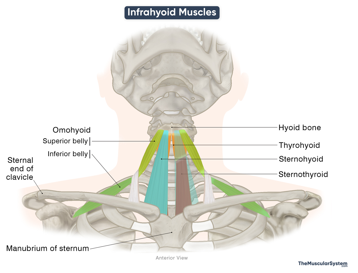
Here are the names of the muscles with their location, anatomy, and primary action:
| Name | Origin | Insertion | Action | Innervation | Blood Supply |
|---|---|---|---|---|---|
| Thyrohyoid Deep layer, on the lateral side | Oblique line on the lamina of the thyroid cartilage | Greater horn (cornu) and the body of the hyoid bone | Elevating the larynx and depressing the hyoid bone to help with swallowing, breathing, and vocalization | Ventral rami of the C1 nerve, via the hypoglossal nerve | Superior laryngeal and infrahyoid branches of the superior thyroid artery |
| Omohyoid Superficial layer, on the lateral side | Inferior belly: Superior border of the scapula Superior belly: The intermediate tendon | Inferior belly: The intermediate tendon, which attaches to the clavicle and first rib by a fibrous sling Superior belly: Inferior border of the body of the hyoid bone | Depressing the hyoid bone and larynx to help resume breathing after swallowing | Ansa cervicalis (C1–C3) | Infrahyoid branches of the superior thyroid artery |
| Sternothyroid* Deep layer, on the medial side | Posterior surface of the manubrium and first costal cartilage | The oblique line of the thyroid cartilage | Depressing the hyoid bone and larynx to help with breathing and producing low-pitched sounds | Ansa cervicalis (C1–C3) | Infrahyoid branch of the superior thyroid artery |
| Sternohyoid Superficial layer, on the medial side | Posterior surface of the sternal end of the clavicle and the manubrium | Inferior border of the body of the hyoid bone | Depressing the hyoid bone and larynx to help resume breathing after swallowing | Ansa cervicalis (C1–C3) | Infrahyoid branch of the superior thyroid artery |
*Note that the sternothyroid is the only muscle in the group that does not have a direct attachment to the hyoid bone.
As evident from the table, all four infrahyoids primarily act to lower the hyoid bone and larynx. During swallowing, the suprahyoid muscles elevate the hyoid and help close the airway; once this phase is complete, the infrahyoid muscles restore the hyoid and larynx to their original position and reopen the airway. This coordinated control allows smooth swallowing, balanced laryngeal movement, and efficient breathing and speech.
Mnemonic
A useful mnemonic to memorize the four muscles’ names is “TOSS.”
- T: Thyrohyoid
- O: Omohyoid
- S: Sternothyroid
- S: Sternohyoid
The infrahyoid muscles, along with the sternocleidomastoid, a major cervical muscle on the lateral neck, form the borders of the thyroid gland. As a result, they serve as key landmarks during medical and surgical procedures.
References
- Infrahyoid Muscles: Radiopaedia.org
- The Infrahyoid Muscles: TeachMeAnatomy.info
- Infrahyoid Muscles: Kenhub.com
- Infrahyoid Muscles: Sciencedirect.com
Della Barnes, an MS Anatomy graduate, blends medical research with accessible writing, simplifying complex anatomy for a better understanding and appreciation of human anatomy.
- Latest Posts by Della Barnes, MS Anatomy
-
Tensor Tympani
- -
Stapedius
- -
Auricularis Posterior
- All Posts


