

Sternocleidomastoid
By
Della Barnes, an MS Anatomy graduate, blends medical research with accessible writing, simplifying complex anatomy for a better understanding and appreciation of human anatomy.
Last updated:
18/11/2025Della Barnes, MS Anatomy
UX/UI Designer at - AdobeDella Barnes, an MS Anatomy graduate, blends medical research with accessible writing, simplifying complex anatomy for a better understanding and appreciation of human anatomy.
The sternocleidomastoid, or SCM, is one of the largest cervical muscles, located on both sides of the neck. It is one of the two superficial neck muscles, the other being the platysma. The functions of the SCM include rotating and flexing the neck.
When you turn your head to one side, the SCM muscle on the opposite side becomes prominent as a ridge running from behind your ear to the top of your chest, making it easy to feel the muscle under the skin.
Anatomy
Location and Attachments
| Origin | Sternal head: Manubrium of sternum Clavicular head: Medial 1/3rd of the clavicle |
| Insertion | Mastoid process of the temporal bone |
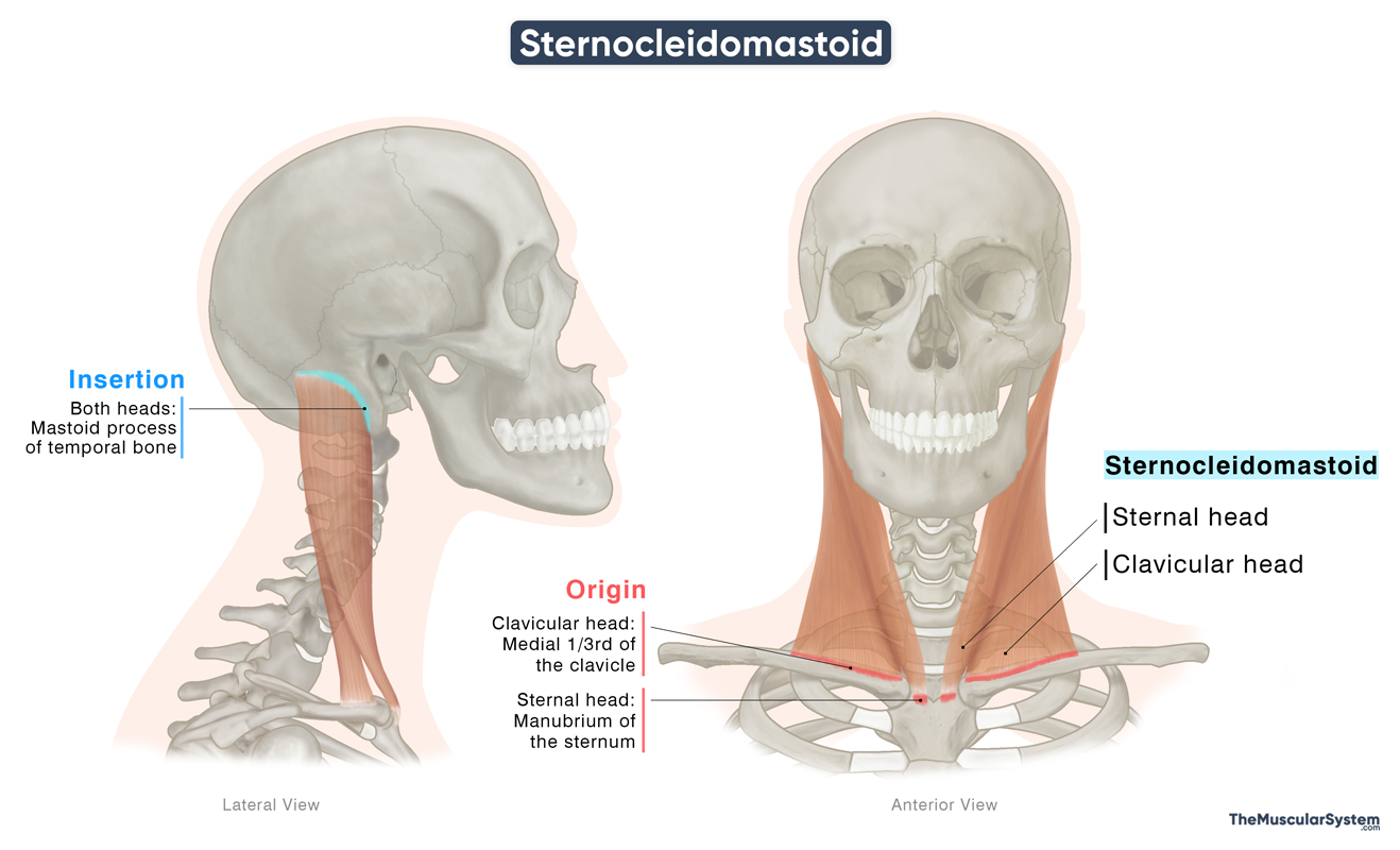
Origin
The muscle originates as two muscular bands from the upper chest, forming two distinct heads named for their points of origin:
- The sternal head arises as a tendinous band from the anterior surface of the manubrium, the upper segment of the sternum.
- The clavicular head is fleshier, originating from the superior anterior surface of the medial one-third of the clavicle (collarbone).
Insertion
The narrow tendinous fibers of the sternal head become fleshy to form a muscular belly that runs upward and obliquely toward the back. The clavicular head also forms a fleshy belly, but it ascends more vertically. The two heads merge to form a single muscle belly, which continues upward to insert into the mastoid process of the temporal bone, just behind the ear, by a narrow yet strong aponeurosis.
The name “sternocleidomastoid” comes from its points of attachment at the sternum, clavicle, and mastoid process.
Relations With Surrounding Muscles and Structures
The muscle is covered by the investing layer of the deep cervical fascia. The platysma, the most superficial muscle in the anterior neck, lies superficial to the SCM, just beneath the skin. Between these two muscles run the external jugular vein and two cutaneous branches of the cervical plexus, the greater auricular and transverse cervical nerves. The parotid gland also lies superficial to the upper part of the SCM.
It is an important landmark, dividing it into two major regions:
- Anterior triangle: Located in front of the SCM, bounded by the muscle’s anterior border, the lower border of the mandible, and the midline of the neck. It contains the suprahyoid and infrahyoid muscles, which assist in swallowing and the movement of the hyoid bone.
- Posterior triangle: Situated behind the SCM, bounded by its posterior border, the anterior border of the trapezius, and the clavicle, which forms the base. It contains the scalene muscles along with several nerves and blood vessels.
At the lower end of the muscle, the sternal and clavicular heads are separated by a small triangular gap called Sedillot’s triangle, part of the larger anterior triangle.
Function
| Action | Unilateral contraction: Bending the neck and rotating the head to one side Bilateral contraction: Bending the neck to the front and lifting the head up |
The muscle plays a vital role in flexing and rotating the cervical vertebra, which rotates the head and bends the neck.
Bilateral Contraction
When the muscle contracts on both sides, it:
- Flexes the lower cervical column to flex the neck, which means lowering your head to bring the chin closer to your chest.
- Extends the atlanto-occipital joint between the cervical column and the base of the skull to lift the head up.
Additionally, it works as an accessory muscle of respiration as it helps elevate the sternum and clavicle during deep breathing.
Unilateral Contraction
When the muscle contracts only on one side, it:
- Causes ipsilateral flexion, which means bending the neck to the same side.
- Rotates the head to the opposite side.
So, when you tilt your head or turn your head to the right, the left SCM muscle is active.
Antagonists
The primary antagonists of the sternocleidomastoid are the splenius capitis and splenius cervicis muscles, because they produce opposite movements, extending the head from a bowed position and bending and rotating it toward the same side.
Innervation
| Nerve | Accessory nerve (CN XI), cervical plexus (C2–C3) |
Motor innervation to the muscle comes from the accessory nerve (cranial nerve XI). It receives sensory and proprioceptive innervation from the cervical plexus, primarily through the anterior rami of the second and third cervical nerves (C2–C3).
Blood Supply
| Artery | Superior thyroid artery |
Blood supply to the muscle comes from the superior thyroid artery, which branches off from the external carotid artery, a major artery of the head and neck regions.
References
- Sternocleidomastoid Muscle: Kenhub.com
- Sternocleidomastoid: Meddean.LUC.edu
- Anatomy, Head and Neck, Sternocleidomastoid Muscle: NCBI.NLM.NIH.gov
- Sternocleidomastoid (SCM) Muscle: Clevelandclinic.org
- Sternocleidomastoid Muscle (Left): Elsevier.com
Della Barnes, an MS Anatomy graduate, blends medical research with accessible writing, simplifying complex anatomy for a better understanding and appreciation of human anatomy.
- Latest Posts by Della Barnes, MS Anatomy
-
Tensor Tympani
- -
Stapedius
- -
Auricularis Posterior
- All Posts


