

Extensor Hallucis Longus
By
Della Barnes, an MS Anatomy graduate, blends medical research with accessible writing, simplifying complex anatomy for a better understanding and appreciation of human anatomy.
Last updated:
04/09/2025Della Barnes, MS Anatomy
UX/UI Designer at - AdobeDella Barnes, an MS Anatomy graduate, blends medical research with accessible writing, simplifying complex anatomy for a better understanding and appreciation of human anatomy.
What is the Extensor Hallucis Longus
The extensor hallucis longus is a long, narrow, unipennate muscle located at the front of the lower leg, along the shin. It is one of the muscles forming the anterior compartment of the lower leg, with the others being the tibialis anterior, extensor digitorum longus, and fibularis tertius muscles.
This muscle plays an important role in foot and toe movement. It extends the big toe and assists in dorsiflexion of the foot, movements that are essential for walking, running, and maintaining balance.
Anatomy
Location and Attachments
| Origin | The middle one-third of the anterior surface of the fibula, and the adjacent interosseous membrane |
| Insertion | Base of the distal phalanx of the hallux (big toe) |
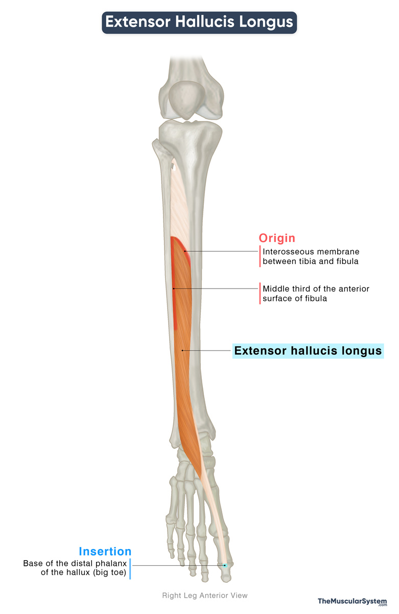
Origin
The tendon of this muscle originates from a long, narrow area on the anterior surface of the fibula, specifically along its middle third. The origin also extends onto the adjacent interosseous membrane between the fibula and tibia.
Insertion
From its origin, the muscle fibers descend medially and thicken to form the muscle belly. Near the distal fibula, the belly tapers into a tendon that continues along the dorsum of the foot. The tendon ultimately inserts onto the dorsal aspect of the base of the distal phalanx of the hallux, commonly known as the big toe.
The long tendon of the muscle is usually visible at the ankle and along the top of the foot, where it stands out most clearly when the big toe is extended.
Relations With Surrounding Muscles and Structures
The extensor hallucis longus (EHL) sits in the middle of the anterior compartment of the leg. It is positioned between the tibialis anterior, which lies medially and slightly in front, and the extensor digitorum longus, which lies on its lateral side.
The anterior tibial artery and vein, along with the deep fibular nerve, pass between the EHL and tibialis anterior. This space serves as a reliable landmark for locating the neurovascular bundle of the lower leg and foot.
As it travels down the leg, the EHL tendon passes under the inferior extensor retinaculum, which holds it in place, preventing it from pulling away from the bone during movement (bowstringing). On the dorsum of the foot, the EHL tendon lies immediately medial to the extensor digitorum longus tendons.
Function
| Action | Extending the great toe and dorsiflexing the ankle joint |
The primary function of the extensor hallucis longus is to extend the hallux, or big toe, both at its metatarsophalangeal and interphalangeal joints. This action makes the functioning of the muscle crucial for fundamental movements such as walking and running.
It helps the tibial anteriors and the other anterior compartment muscles to dorsiflex the foot at the ankle joint, which means extending the foot toward the shin.
The muscle also weakly assists with the inversion or inward rotation of the foot, and contributes to stability during standing and walking.
Antagonists
The flexor hallucis longus, located in the deep posterior compartment of the leg, and the flexor hallucis brevis, located in the foot, both flex the big toe. Their action opposes that of the extensor hallucis longus, making them its antagonists.
Innervation
| Nerve | Deep fibular nerve (L5-S1) |
It receives innervation from the deep fibular nerve, which arises from the fifth (sometimes fourth) lumbar and first sacral nerve roots. This nerve is a branch of the common fibular nerve, itself derived from the sciatic nerve.
Blood Supply
| Artery | Anterior tibial artery |
The muscle primarily gets its blood supply from the anterior tibial artery and its branches. Supplementary blood supply comes from the perforating branch of the fibular artery.
References
- Anatomy, Bony Pelvis and Lower Limb: Extensor Hallucis Longus Muscle: NCBI.NLM.NIH.gov
- Extensor Hallucis Longus Muscle: Elsevier.com
- Extensor Hallucis Longus Muscle: Kenhub.com
- Extensor Hallucis Longus Muscle: Radiopaedia.org
Della Barnes, an MS Anatomy graduate, blends medical research with accessible writing, simplifying complex anatomy for a better understanding and appreciation of human anatomy.
- Latest Posts by Della Barnes, MS Anatomy
-
Tensor Tympani
- -
Stapedius
- -
Auricularis Posterior
- All Posts


