

Biceps Femoris
By
Della Barnes, an MS Anatomy graduate, blends medical research with accessible writing, simplifying complex anatomy for a better understanding and appreciation of human anatomy.
Last updated:
22/08/2025Della Barnes, MS Anatomy
UX/UI Designer at - AdobeDella Barnes, an MS Anatomy graduate, blends medical research with accessible writing, simplifying complex anatomy for a better understanding and appreciation of human anatomy.
What is the Biceps Femoris
The biceps femoris is a long, fusiform muscle located along the back of the thigh. It belongs to the posterior compartment of thigh muscles, and is part of the Hamstring muscle group along with semitendinosus and semimembranosus.
The muscle gets its name from its two-headed structure. The long head participates in movements at both the hip and knee joints, while the short head only works on the knee.
Anatomy
Location and Attachments
The two heads of the biceps femoris originate from completely different points, as one rises from the hip bone, while the other rises from the femur or thigh bone. The two heads have a common point of insertion.
| Origin | — The ischial tuberosity (long head) — The linea aspera and the lateral supracondylar line of the femur (short head) |
| Insertion | The lateral surface of the head of fibula, and the lateral condyle of tibia |
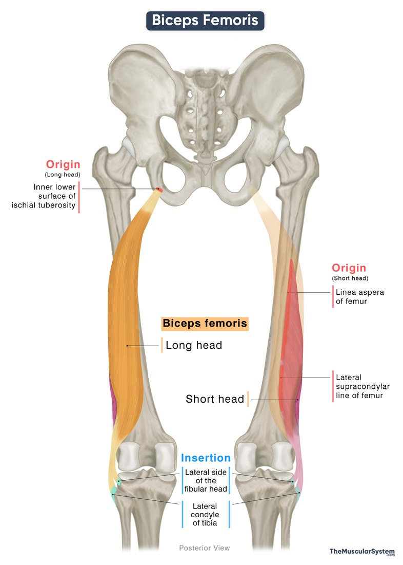
Origin
Long Head
The long head of the biceps femoris originates from the inner and lower part of the posterior ischial tuberosity, commonly known as the “sit bone.” It shares this origin with the semitendinosus muscle, which arises slightly more laterally. This region also serves as the attachment point for the sacrotuberous ligament.
Short Head
The short head has a relatively broad origin, arising from the lower third of the lateral lip (or outer edge) of the linea aspera of the femur. The point of origin extends down to the lateral supracondylar ridge.
Because it originates from the femur rather than the ischium, the short head of the biceps femoris is often excluded from the hamstring group, with only the long head considered a true hamstring muscle.
Biceps Femoris Insertion
From their origin at the ischium, the tendons of the long head of biceps femoris and semitendinosus descend together briefly before dividing into their respective muscle bellies. The long head of the biceps femoris forms a fleshy, fusiform belly that travels obliquely and laterally down the thigh.
As it approaches the distal femur, the long head transitions into an aponeurosis. Here, it merges with the fibers of the short head of the biceps femoris, forming a common tendon. This conjoined tendon then bifurcates slightly and becomes closely associated with the lateral collateral ligament of the knee.
Ultimately, the tendon inserts into the lateral surface of the proximal end or head of the fibula (the shin bone). Additionally, a smaller portion of the tendon extends to insert on the lateral condyle of tibia.
Relations With Surrounding Muscles and Structures
The biceps femoris is the most lateral and superficial muscle of the posterior thigh, covered only by skin and fascia along most of its length. The proximal end of the long head is covered posteriorly by the gluteus maximus.
Medial to the biceps femoris lie the semitendinosus and semimembranosus muscles. The semimembranosus also lies deep to this muscle, along with the adductor magnus from the thigh’s medial compartment. Inferiorly, near the knee joint, the lateral head of the gastrocnemius lies deep to the distal portion of the muscle.
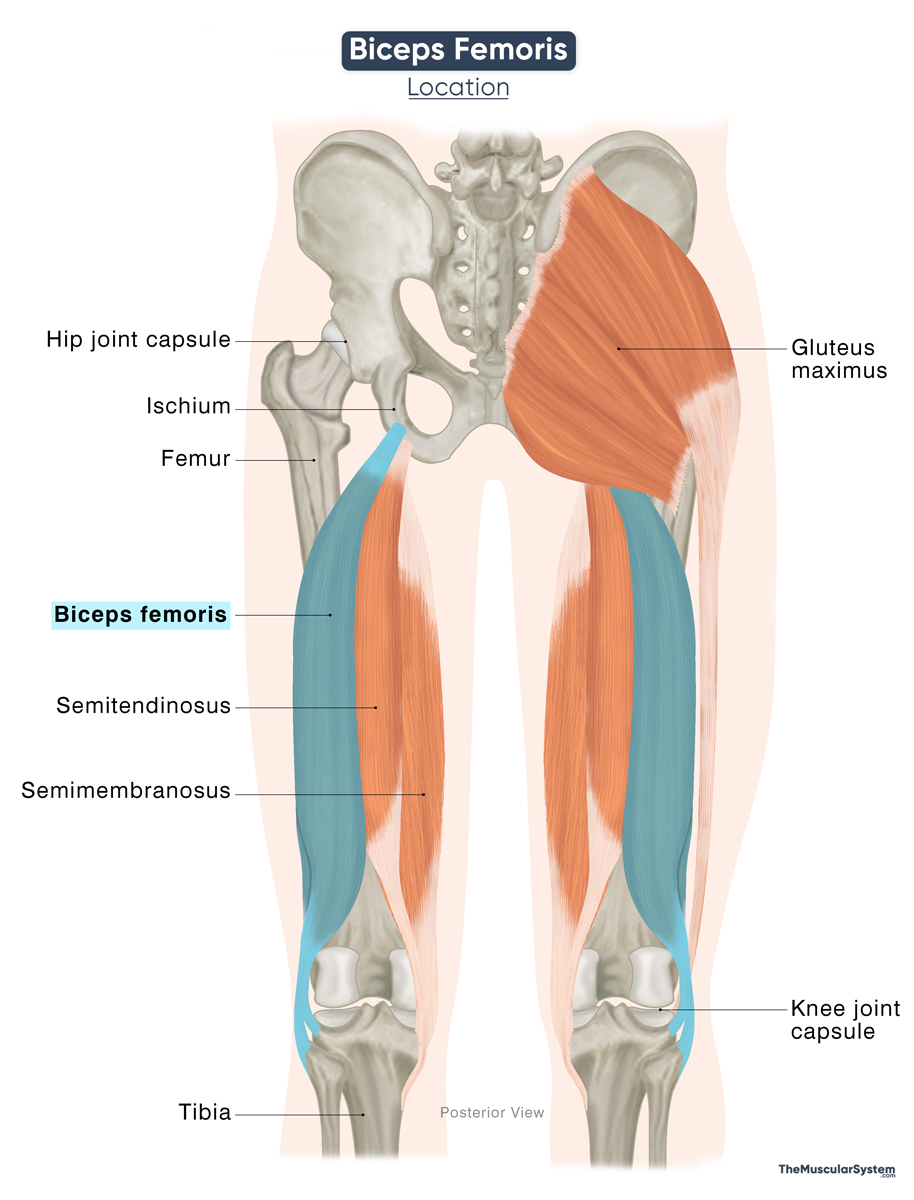
The biceps femoris forms the lateral boundary of the popliteal fossa, a triangular depression at the back of the knee through which major nerves and blood vessels pass into the leg.
The sciatic nerve runs deep to the biceps femoris in the posterior thigh. Laterally, the iliotibial (IT) tract courses in close proximity along the muscle’s border.
Biceps Femoris Function
| Action | — Flexing and laterally rotating the knee joint (both heads) — Extending the hip joint (only the long head) |
Having attachments at the hip and the knee joint, the long head of the biceps femoris contributes to both flexion of the lower leg at the knee joint and extension of the thigh at the hip joint. So, it helps with both folding the leg at the knee joint, like when sitting cross-legged, and straightening the hip joint, like when standing up from a chair.
Being active at both ends, the long head is a weak knee flexor when the hip is already extended, and vice versa (active insufficiency).
The short head, attached only at the knee joint, plays a vital role in flexing the knee.
Both heads together help rotate the leg laterally at the knee joint, which involves rotating your lower leg outwards, such as when getting into a car.
Antagonists
Since the primary function of the muscle is to flex the knee joint, its antagonists are the quadriceps muscles, the rectus femoris, and the three vastus muscles, as they function to extend the knee.
Innervation
| Nerve | — Tibial nerve (L5-S2) for the long head — Common fibular nerve (L4-S3) for the short head |
The long head of the biceps femoris, which functions as a knee flexor, is supplied by the tibial division of the sciatic nerve, originating from the fifth lumbar and second sacral nerve roots (L5-S2).
The short head is innervated by the common fibular (peroneal) division of the sciatic nerve, also from the fourth lumbar to third sacral nerve roots (L4-S3).
Blood Supply
| Artery | Perforating, inferior gluteal, and popliteal arteries |
The biceps femoris muscle receives its primary blood supply from the perforating branches of the deep femoral artery (profunda femoris) and the muscular branches of the popliteal artery. The deep femoral artery is a major branch of the femoral artery, while the popliteal artery is a continuation of the femoral artery after it passes through the adductor hiatus.
The inferior gluteal artery, a branch of the internal iliac artery, also contributes to the muscle’s vasculature.
Supplementary vascular supply comes from the lateral superior genicular artery, a branch of the popliteal artery, and the medial circumflex femoral artery, a branch of the deep femoral artery.
References
- Biceps Femoris: Osmosis.org
- Biceps Femoris Muscle: Radiopaedia.org
- Biceps Femoris: Origin, Insertion, Action, and Innervation: GetBodySmart.com
- Biceps Femoris Muscle: Kenhub.com
- Biceps Femoris Muscle (Anatomy): PrimaryCareNotebook.com
- Biceps Femoris Muscle: IMAIOS.com
- Long Head of Biceps Femoris: Elsevier.com
- Short Head of Biceps Femoris: Elsevier.com
Della Barnes, an MS Anatomy graduate, blends medical research with accessible writing, simplifying complex anatomy for a better understanding and appreciation of human anatomy.
- Latest Posts by Della Barnes, MS Anatomy
-
Tensor Tympani
- -
Stapedius
- -
Auricularis Posterior
- All Posts


