Intertransversarii
Last updated:
06/01/2026Della Barnes, an MS Anatomy graduate, blends medical research with accessible writing, simplifying complex anatomy for a better understanding and appreciation of human anatomy.
What Are the Intertransversarii
Intertransversarii are small paired muscles located deep in the human back. They form the deepest layer of the intrinsic back muscle group, along with the interspinales. The tiny muscles are located on either side of the spine, between the vertebra, and play a vital role in stabilizing the vertebral column.
Anatomy
Location and Attachments
Since the muscles span the entire length of the spinal column, they are divided into groups based on their points of origin and insertion, with the cervical and lumbar parts being the most well-developed.
| Origin | Posterior and anterior tubercles of the transverse processes of the cervical vertebra; the transverse and accessory processes of the lumbar vertebra. |
| Insertion | The transverse processes and mammillary processes (in the cervical region) of the vertebrae inferior to the point of origin |
Intertransversarii Cervicis (or Colli)
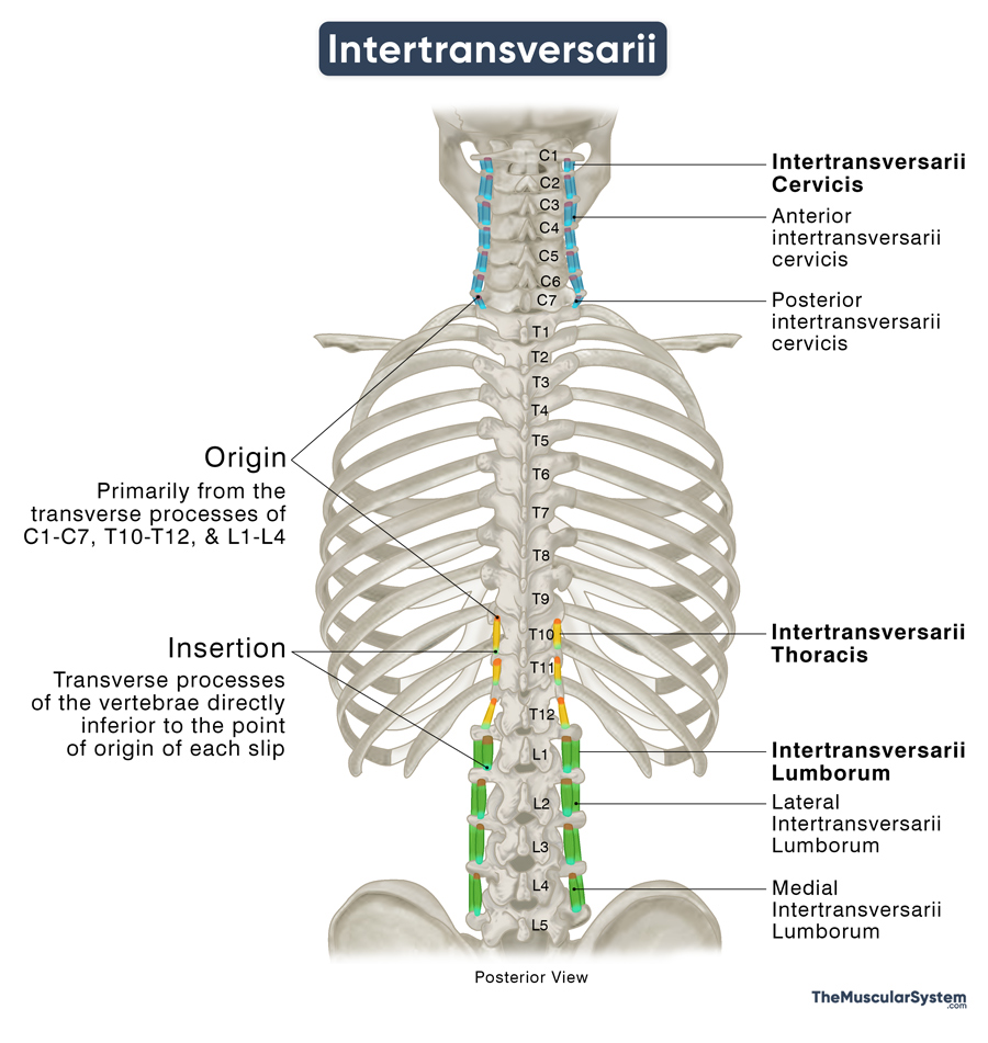
The intertransversarii cervicis muscles are the most well-defined part of the transversospinales muscle group. It consists of 7 pairs of small muscular slips or fascicles, each having an anterior and posterior component. They originate from the superior surfaces of the anterior and posterior tubercles of the transverse processes of all 7 cervical vertebrae.
- Anterior intertransversarii cervicis: The slips originating from the anterior tubercles
- Posterior intertransversarii cervicis: The slips originating from the posterior tubercles
The muscle slips descend to insert into the transverse processes of the cervical vertebrae directly inferior to their points of origin. Specifically, the anterior intertransversarii cervicis is inserted into the anterior tubercle of the next cervical vertebra, while the posterior intertransversarii cervicis attach to the posterior tubercles.
For a clearer picture of their orientation, the slips originating from the 1st cervical vertebra (atlas) insert into the 2nd cervical vertebra (axis), while those from the 7th cervical vertebra (C7) insert into the 1st thoracic vertebra (T1).
Intertransversarii Thoracis
The muscle is the least developed in the thoracic region, and is often completely absent. When present, the muscle slips may be found between the last cervical vertebra (C7) and the first thoracic vertebra (T1), as well as between the lower thoracic vertebrae (specifically T10 to T12) and between the last thoracic vertebra (T12) and the first lumbar vertebra (L1). Consequently, the thoracic intertransversarii are often considered extensions of the cervical and lumbar parts of the muscle rather than a distinct region on their own.
Intertransversarii Lumborum
As indicated by the name, the intertransversarii lumborum constitutes the lumbar part of the muscle and is reasonably well-developed. There are 4 pairs of muscular slips in this region, each comprising one lateral and one medial component. These slips originate from the transverse and accessory processes of the 1st to 4th lumbar vertebrae (L1-L4).
- Lateral Intertransversarii Lumborum: The fascicles originating from both transverse and accessory processes
- Medial Intertransversarii Lumborum: The fascicles originating solely from the accessory processes
The muscle fibers descend, with the lateral component inserting into the transverse process of the subsequent lumbar vertebra, while the medial component inserts into the mammillary process. Consequently, each pair of muscular slips is located between consecutive lumbar vertebrae: the first pair between the first and second lumbar vertebrae (L1-L2), the second pair between L2 and L3, and so on, until L4 and L5.
Relations With Surrounding Muscles and Structures
Since it is the deepest layer of the intrinsic back muscles, it has the interspinales muscles lying superior to it. Another point worth noting is that the anterior intertransversarii cervicis are located deep to the posterior intertransversarii cervicis.
In the cervical or neck area, the intertransversarii cervicis muscles have the scalene muscles lying laterally or on their sides, while the longus colli lie in front.
In the lumbar region, the multifidus and rotatores longus muscles lie superior to the intertransversarii. The lateral intertransversarii lumborum are positioned posterior to the iliopsoas, which is part of the deep muscle group in the iliac region. Laterally, the intertransversarii are bordered by the quadratus lumborum, a deep abdominal muscle, along with the intertransverse ligaments.
Function
| Action | Stabilizing the spinal column and assisting in lateral flexion of cervical and lumbar spines |
The functions of intertransversarii are still unclear. According to medical literature, the small size of the muscles makes them unsuitable for initiating any significant spinal movement. Instead, they are more important for maintaining spinal stability and flexibility through bilateral contraction. Their high density of muscle spindles suggests a key role in spinal proprioception, which helps the body sense the position and movement of the spine and trunk.
Additionally, during unilateral or one-sided contraction, these muscles are believed to assist the flexors of the neck and the lumbar spine to laterally flex the body trunk on the same side as the contracting muscles.
Innervation
| Nerve | Anterior and posterior rami of corresponding spinal nerves |
In the cervical region, the anterior cervical intertransversarii are supplied by the anterior (ventral) rami of the 1st to 7th cervical spinal nerves, while the posterior cervical intertransversarii are innervated by both the anterior and posterior (dorsal) rami of the same nerves.
In the lumbar region, both the lateral and medial intertransversarii lumborum receive innervation from the anterior rami of the 1st to 4th lumbar spinal nerves.
Blood Supply
| Artery | Deep cervical, occipital, vertebral, and lumbar arteries |
The cervical part, mainly the anterior cervical intertransversarii, is supplied by the deep cervical, occipital, and vertebral arteries, branching from the costocervical trunk, external carotid artery, and subclavian artery, respectively. The posterior cervical muscle slips receive blood supply from the ascending cervical artery, a branch of the inferior thyroid artery.
The lumbar part gets its primary blood supply from the lumbar artery, particularly its dorsal branches.
References
- Intertransversarii: TeachMeAnatomy.info
- Intertransversarii Muscle Group: Radiopaedia.org
- Intertransversarii Lumborum Muscles: Kenhub.com
- Anterior Intertransversarii Colli Muscles: Kenhub.com
- Posterior Intertransversarii Colli Muscles: Kenhub.com
- Lateral Intertransversarii Lumborum Muscles (Right): Elsevier.com
Della Barnes, an MS Anatomy graduate, blends medical research with accessible writing, simplifying complex anatomy for a better understanding and appreciation of human anatomy.
- Latest Posts by Della Barnes, MS Anatomy
-
Corrugator Supercilii
- -
Depressor Supercilii
- -
Orbicularis Oculi
- All Posts


