Transversospinalis Muscles
Last updated:
30/10/2024Della Barnes, an MS Anatomy graduate, blends medical research with accessible writing, simplifying complex anatomy for a better understanding and appreciation of human anatomy.
What Are the Transversospinalis
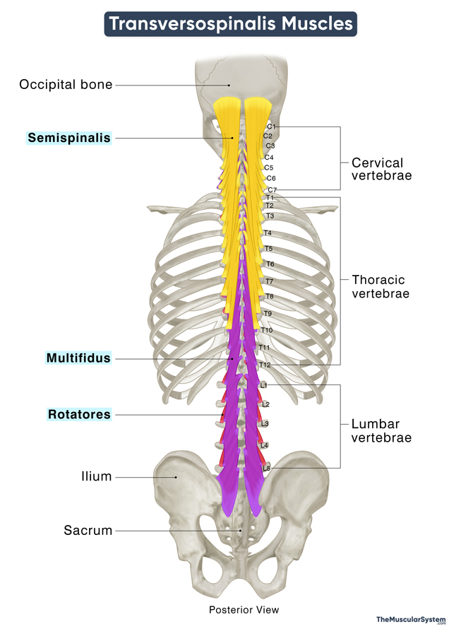
The transversospinalis muscles, collectively referred to as the transversospinales, form the deep layer of intrinsic back muscles. They connect the vertebrae to each other and, in some cases, to the skull, playing a key role in stabilizing the vertebral column.
Names and Basic Anatomy of the Transversospinales Muscles
The group includes 3 muscles, each consisting of small fascicles, or muscle strips, located along the sides of the vertebral column. Each muscle can be divided into regions based on the origin of these muscle strips along the cervical, thoracic, or lumbar vertebrae.
The name “transversospinales” is derived from the fact that most of these fascicles originate from the transverse processes of the vertebrae and are inserted into the spinous process. Here is a snapshot of the muscles belonging to this group:
| Name | Origin | Insertion | Action | Innervation | Blood Supply |
|---|---|---|---|---|---|
| Semispinalis (Most superficial of the 3) | Articular processes of the cervical vertebrae (for semispinalis capitis) and transverse processes of the cervical, thoracic, and lumbar vertebrae | The occipital bone and the spinous processes of the cervical and thoracic vertebrae | Extending, laterally bending, and rotating the head, neck, and back (specific to semispinalis thoracis) | Dorsal rami of cervical and thoracic nerves | Occipital, vertebral, deep cervical, and posterior intercostal (dorsal rami) arteries |
| Multifidus (Lies between the other 2) | Articular processes of C4 to C7 vertebrae; transverse processes of all thoracic vertebrae; mammillary processes of all lumbar vertebrae; dorsal side of the sacrum and ilium | The occipital bone and the spinous processes of the cervical and thoracic vertebrae spanning | Stabilizing, extending, laterally flexing, and rotating the spine | Dorsal rami of the corresponding spinal nerves | Occipital, deep cervical, vertebral, subcostal, posterior intercostal, lumbar, and lateral sacral arteries |
| Rotatores (Deepest of the 3) | Transverse processes of all the cervical, thoracic, and lumbar vertebrae, with the thoracic part being the most prominent | Spinous process and laminae of vertebrae located 1-2 vertebral segments above the origin of each muscular strip | Stabilizing the vertebral column, straightening and rotating the thoracic spine | Dorsal rami of the spinal nerves corresponding to each muscle strip | Posterior/dorsal branches of posterior intercostal and lumbar arteries |
As the intermediate layer of the intrinsic back muscles, the transversospinalis muscles lie deep to the splenius muscles from the superficial layer and the erector spinae from the intermediate layer.
The position of the transversospinalis muscles on the vertebral column is such that they get little leverage to move the spine. Still, they help the splenius and erector spinae to extend, flex, and rotate the spine. However, as mentioned above, their primary function remains to keep the spine stable and flexible by maintaining a connection between the vertebrae during various movements.
The multifidus is the longest of the three muscles, spanning the entire length of the vertebral column, from the cervical vertebrae to the sacrum and ilium.
References
- Transversospinal Muscles (Left): Elsevier.com
- Transversospinal Muscles: IMAIOS.com
- Transversospinal Muscle Group: Radiopaedia.org
- Deep Back Muscles: Kenhub.com
Della Barnes, an MS Anatomy graduate, blends medical research with accessible writing, simplifying complex anatomy for a better understanding and appreciation of human anatomy.
- Latest Posts by Della Barnes, MS Anatomy
-
Flexor Digiti Minimi Brevis of the Foot
- -
Adductor Hallucis
- -
Flexor Hallucis Brevis
- All Posts


