Chest Muscles
While the pectoralis major and minor are often the most recognized muscles in the chest area, they represent only a part of the complex muscular structure in this region. The chest comprises several other vital components, including the intercostal muscles and associated structures essential for the movement and support of the ribcage, shoulders, and arms.
Names of All the Muscles in the Chest With Location & Anatomy
The muscles in this group are all attached to the axial skeleton (skull, ribcage, vertebral column, and sternum), as they all originate from the ribs, vertebrae, or sternum.
Muscles in the Thoracic Region
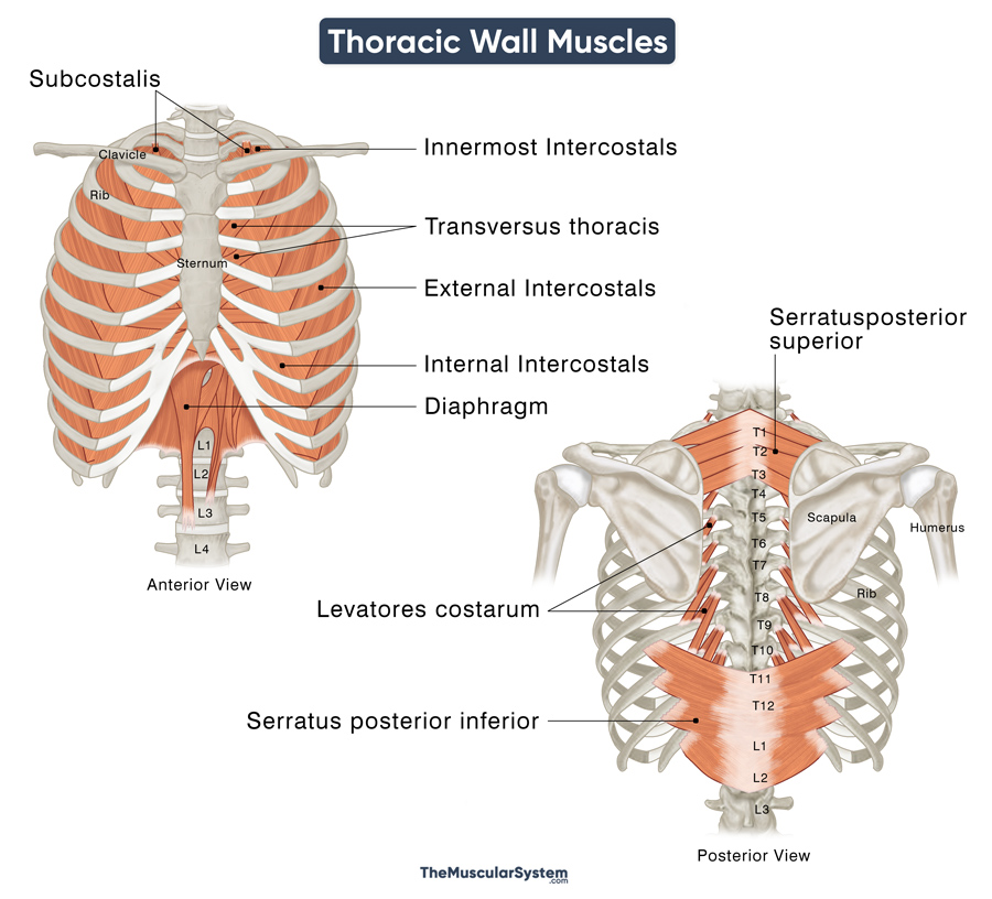
Some chest muscles, like the intercostals, are considered axial muscles, as their origin and insertion both lie in the axial skeleton. These muscles, which lie deep in the thoracic region, form the walls of the thoracic cavity and mostly help with respiration.
Here is a list of all the muscles in the thoracic region:
| Name | Location | Attachments | Function |
|---|---|---|---|
Thoracic (Chest) Wall Muscles |
|||
| Intercostals | Intercostal spaces between ribs | Origin: Ribs Insertion: Ribs |
Elevating and depressing the ribs to control thoracic volume during breathing |
| — External | Between the ribs, from the spines in the back to the sternum at the front, slanting downward and forward from the rib above to the rib below. | Origin: Lower borders of 1st to 11th ribs Insertion: Upper borders of 2nd-12th ribs |
Elevating the ribcage to increase its volume during forced inhalation |
| — Internal | Occupy the same space between the ribs but lie beneath the external intercostals. They run in the opposite direction, slanting downward and backward. | Origin: Costal groove of 1st to 11th ribs Insertion: Upper borders of 2nd-12th ribs |
Depressing the rib cage to reduce its volume during forced exhalation |
| — Innermost | Deep to the internal intercostals, occupying the same spaces and having the same orientation of the muscle fibers. | Origin: Interior surface and borders of the 1st-11th ribs Insertion: Upper borders of 2nd-12th ribs |
Maintaining the integrity of the ribcage during respiration and helping the internal intercostals |
| Subcostalis | One of the deepest muscles in thoracic region, most prominent along the inner surface of the lower ribs, often spanning multiple intercostal spaces. | Origin: Angle of the ribs, especially the lower ribs Insertion: Inner surface and upper borders of the ribs below their origin |
Depressing the rib cage during forced exhalation |
| Transversus thoracis | On the inside of the front of the rib cage, right behind the sternum and the costal cartilages. | Origin: Posterior surface of the xiphoid process and body of sternum, and costal cartilages of 4th-7th ribs. Insertion: Costal cartilage of 2nd-6th ribs |
Depressing the ribs (mainly 2nd-6th) during forced exhalation |
| Levatores costarum | Along the back, running from the spine to ribs at the back of the ribcage. | Origin: Transverse processes of C7-T11 vertebrae Insertion: Upper surface of the rib immediately below the origin of each strip |
Elevating the ribcage during inhalation |
| Serratus posterior | Group of muscles connecting the vertebrae to the ribs. | Origin: Spinous processes of vertebrae Insertion: Ribs |
Helping with respiration |
| — Serratus posterior inferior | Lower back region, below the shoulder blades, and above the hips. | Origin: Spinous processes of T11-L2 vertebrae Insertion: Lower borders of 9th-12th ribs |
Depressing the lower ribs during exhalation |
| — Serratus posterior superior | Upper back region, just beneath the neck, and between the shoulder blades. | Origin: Nuchal ligament and spinous processes of the C7-T3 vertebrae Insertion: Upper borders of 2nd-5th ribs |
Elevating the upper ribs during inhalation |
Other Muscles in the Thoracic Cavity |
|||
| Diaphragm | Dome-shaped sheet muscle between the thoracic and abdominal cavities, separating the two. | Origin: In three parts, from the back of the xiphoid process, inner surface of 7th-12th ribs, and 1st-3rd lumbar vertebrae Insertion: Central tendon of the diaphragm |
A primary muscle of respiration, helping in increasing and decreasing the thoracic volume to draw air in and then expel it for breathing to happen |
Muscles in the Pectoral Region
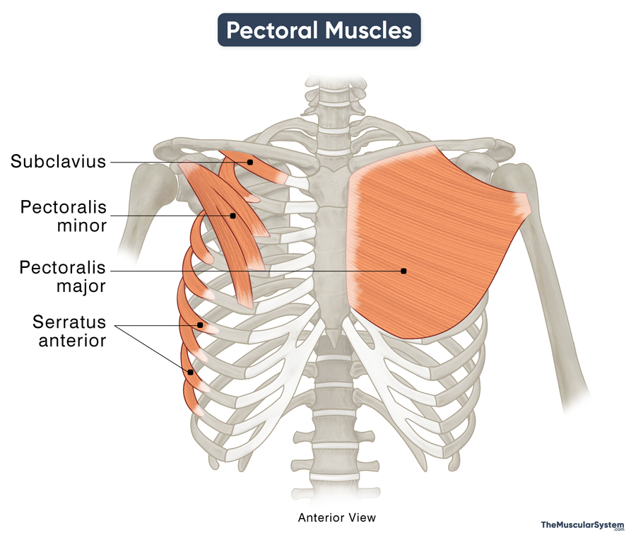
Muscles like the pectoralis major and minor attach to both the axial and appendicular skeletons, making them axioappendicular muscles. Their actions often involve helping shoulder and arm movements. These lie superficially in the chest and are grouped into the muscles of the pectoral region.
| Name | Location | Attachments | Function |
|---|---|---|---|
| Pectoralis major | The biggest chest muscle, extending from the shoulder region across the front of the chest, covering the ribs. | Origin: In two heads, from the clavicle and the costal cartilages of the 1st-6th ribs. Insertion: Bicipital groove and greater tubercle of the humerus |
Flexing the arms forward, rotating them at the shoulder, and adducting them in front, across the chest |
| Pectoralis minor | In the upper chest, near the shoulder, under the pectoralis major | Origin: Sternal end of 3rd-5th ribs Insertion: Coracoid process of scapula |
Stabilizing the scapula, and elevating the 3rd-5th ribs during inhalation |
| Subclavius | At the front of the chest, underneath the collarbones | Origin: Sternal end of 1st rib Insertion: Subclavian groove of the clavicle or collarbone |
Depressing the clavicle and keeping it steady during shoulder movements |
| Serratus anterior | On the sides of the chest, under the armpits, and along the ribs. | Origin: in three parts, from 1st-8th ribs Insertion: In three parts, into the scapula’s angles and borders |
Abducting the scapula and rotating it when raising the arms |
References
- Muscles of the Pectoral Region: TeachMeAnatomy.info
- Anatomy, Shoulder and Upper Limb, Pectoral Muscles: https://www.NCBI.NLM.NIH.gov
- Chest & Abdominal Muscle Groups: Study.com
- The Muscles of the Thoracic Cage: TeachMeAnatomy.info
- Muscles of the thoracic wall: Osmosis.org
- Muscles of Thorax: Elsevier.com


