Rhomboid Major
Last updated:
28/06/2023Della Barnes, an MS Anatomy graduate, blends medical research with accessible writing, simplifying complex anatomy for a better understanding and appreciation of human anatomy.
What is Rhomboid Major
The rhomboid major is a paired superficial muscle located in the upper back of the human body. The major and minor rhomboids are collectively called the rhomboids, which form the superficial layer of extrinsic back muscles with the trapezius, latissimus dorsi, and levator scapulae.
Anatomy
Location and Attachments
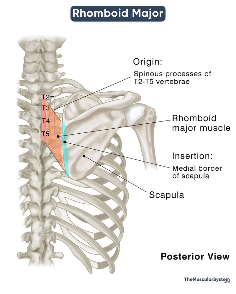
| Origin | Spinous processes of T2 to T5 vertebrae |
| Insertion | The medial border of the scapula |
Origin
Each rhomboid major muscle can individually be described as a broad quadrilateral muscle extending from the thoracic spine to the scapula on the left and right sides of the back.
After originating from the spinous processes of the T2 through T5 vertebrae, the paired muscle courses obliquely in an inferolateral direction on both sides of the back.
Insertion
The muscle fibers run diagonally, converging into a narrow tendon as they reach the scapula’s medial border, where they insert between the bone’s inferior angle and spine. The point of insertion involves both the dorsal and costal facets of the scapula’s medial border.
Relations With Surrounding Muscles and Structures
The relatively small muscle is covered entirely by the trapezoid, the only muscle lying superficial to it. The one point where the rhomboid major lies unobstructed is the auscultation triangle, an anatomical space in the mid-back located over the rhomboid major’s inferior border. The muscle forms the superolateral border of this space. The latissimus dorsi and trapezius form the inferior and superomedial walls, respectively.
The rhomboid major is inferior to the rhomboid minor, with the latter overlapping its superior part. Though the major and minor rhomboids are usually totally separate muscles, they may sometimes be fused partially, displaying one anatomical variation of the muscle.
Function
| Action | Retracting and rotating the scapula at the scapulothoracic joint |
The rhomboid major muscle connects the thoracic spine with the scapula as it extends between the spinous processes and the scapular border. This location is vital to the functioning and the structure of the shoulder and upper arms.
Contraction of this muscle stabilizes the scapula against the ribcage at the scapulothoracic joint. It allows it to rotate and retract towards the spine during shoulder and arm movements, resulting in inferior rotation of the glenoid cavity, letting you drop your shoulders.
The muscle also plays an essential role in stabilizing the shoulder girdle and keeping the scapula in place when the other muscles in the region work to move the shoulder and upper arm.
Antagonists
Serratus anterior, the fan-shaped chest muscle attached to the ribcage, acts as its antagonist.
Innervation
| Nerve | Dorsal scapular nerve (C4, C5) |
Blood Supply
| Artery | Dorsal scapular artery |
Its primary blood supply comes from the dorsal scapular artery that branches from the thyrocervical trunk, which itself is a branch of the subclavian artery.
Reference
- Rhomboid Major Muscle: IMAIOS.com
- Rhomboid Major: TeachMeAnatomy.info
- Rhomboid Major Muscle: GetBodySmart.com
- Rhomboid Major: Meddean.LUC.edu
- Rhomboid Major Muscle: Location, Function, and Structure: Study.com
- Rhomboid Major: HealthLine.com
- Rhomboid Muscles: KenHub.com
Della Barnes, an MS Anatomy graduate, blends medical research with accessible writing, simplifying complex anatomy for a better understanding and appreciation of human anatomy.
- Latest Posts by Della Barnes, MS Anatomy
-
Rectus Capitis Posterior Major
- -
Obliquus Capitis Inferior
- -
Obliquus Capitis Superior
- All Posts


