

Salpingopharyngeus
By
Della Barnes, an MS Anatomy graduate, blends medical research with accessible writing, simplifying complex anatomy for a better understanding and appreciation of human anatomy.
Last updated:
22/12/2025Della Barnes, MS Anatomy
UX/UI Designer at - AdobeDella Barnes, an MS Anatomy graduate, blends medical research with accessible writing, simplifying complex anatomy for a better understanding and appreciation of human anatomy.
What is the Salpingopharyngeus
The salpingopharyngeus is a small, narrow, paired muscle in the pharyngeal wall in the neck. It is one of the three longitudinal pharyngeal muscles, with the other two being the stylopharyngeus and palatopharyngeus. The muscle derives its name from the Greek word sálpinx (“trumpet”), a reference to its long, trumpet-like shape.
The muscle elevates the pharynx to help with both swallowing and speech, and maintains air pressure in the middle ear.
Anatomy
Location and Attachments
| Origin | The inferior cartilaginous portion of the Eustachian tube |
| Insertion | Blends with the palatopharyngeus muscle |
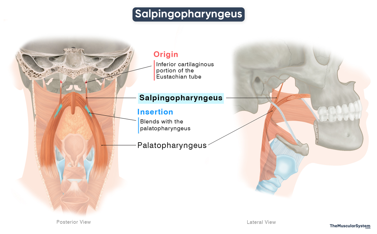
Origin
The muscle originates from the lower end of the cartilaginous portion of the Eustachian (auditory) tube, where it opens into the nasopharynx.
Insertion
From there, the muscle fibers course inferiorly as they form a slender muscle belly, which finally blends into the palatopharyngeus muscle.
Relations With Surrounding Muscles and Structures
The muscle lies posterior to the torus tubarius, a prominence of the nasopharyngeal mucous membrane. From this region, it passes through the salpingopharyngeal fold, a vertical ridge of mucosa visible along the lateral wall of the nasopharynx.
Near its origin, the levator veli palatini, an important muscle in the soft palate, lies behind the salpingopharyngeus. Inferiorly, the muscle does not remain as a distinct structure. As it reaches the back of the lateral border of the soft palate, it blends with the fibers of the palatopharyngeus.
Function
| Action | — Elevating the pharynx and larynx to assist with swallowing and vocalization — Helping open the Eustachian tube to equalize air pressure |
Along with the other longitudinal pharyngeal muscles, the salpingopharyngeus elevates and shortens the pharynx and larynx during swallowing. This elevation helps widen the pharyngeal cavity, allowing the bolus to pass further, as the pharyngeal constrictor muscles propel it toward the esophagus.
By elevating the larynx and pharynx, the muscle also contributes to vocalization by influencing voice resonance. In addition, due to its close anatomical relationship with the auditory tube, the salpingopharyngeus indirectly assists the tensor veli palatini in opening the Eustachian tube during swallowing to equalize air pressure in the middle ear.
Antagonists
It does not have any direct antagonist. Once its action ends, the infrahyoid muscles depress the larynx and pharynx to send them back to their resting positions.
Innervation
| Nerve | Pharyngeal plexus (CN X) |
The muscle is innervated by the pharyngeal plexus, carrying motor fibers from the vagus nerve (CN X)
Blood Supply
| Artery | Ascending palatine artery, greater palatine artery, and pharyngeal trunk of the ascending pharyngeal artery |
Blood supply to the muscle comes from three sources: the ascending palatine artery, which is a branch of the facial artery; the greater palatine artery, a branch of the maxillary artery; and the pharyngeal trunk of the ascending pharyngeal artery.
References
- Salpingopharyngeus: TeachMeAnatomy.info
- Salpingopharyngeus Muscle: Kenhub.com
- Salpingopharyngeus Muscle: Radiopaedia.org
- Salpingopharyngeus Muscle: Elsevier.com
- Salpingopharyngeus: Meddean.LUC.edu
Della Barnes, an MS Anatomy graduate, blends medical research with accessible writing, simplifying complex anatomy for a better understanding and appreciation of human anatomy.
- Latest Posts by Della Barnes, MS Anatomy
-
Tensor Tympani
- -
Stapedius
- -
Auricularis Posterior
- All Posts


