

Hamstring
By
Della Barnes, an MS Anatomy graduate, blends medical research with accessible writing, simplifying complex anatomy for a better understanding and appreciation of human anatomy.
Last updated:
19/08/2025Della Barnes, MS Anatomy
UX/UI Designer at - AdobeDella Barnes, an MS Anatomy graduate, blends medical research with accessible writing, simplifying complex anatomy for a better understanding and appreciation of human anatomy.
What is the Hamstring Muscle Group
The hamstring is a group of three muscles in the posterior compartment of the thigh, located between the hip and knee joints. These muscles play a vital role in the movements of the lower limb.
The term “hamstrings” comes from Old English “ham,” meaning the bend of the knee, and “string,” referring to the tendons at the back of the knee. All three muscles are attached to the knee joint via long, thin tendons.
Characteristics of the Hamstring Muscles
All hamstring muscles have several common structural and functional characteristics, including:
- They originate from the ischial tuberosity
- They cross both the hip and knee joints
- They are innervated by the tibial division of the sciatic nerve
- They insert into the tibia and/or fibula
- Their function includes extending the hip and flexing the knee
Names of the Hamstring Muscles With Location & Anatomy
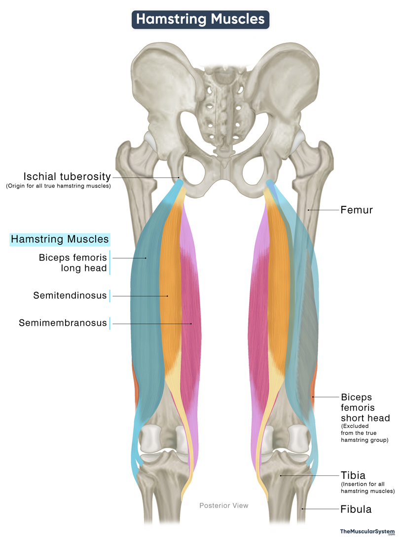
Here is a list of the muscles that make up the hamstring group, along with their locations in the posterior thigh and basic anatomical details. From lateral to medial, the hamstring muscles are:
| Name | Origin | Insertion | Innervation | Blood Supply |
|---|---|---|---|---|
| Biceps femoris | ||||
| — Long head | Lower inner surface of the ischial tuberosity | — Lateral surface of the proximal fibula — Lateral tibial condyle | Tibial nerve (L5-S2) | Perforating, inferior gluteal, and popliteal arteries |
| — Short head | Linea aspera, and lateral supracondylar line of the femur | Same as the long head | Common fibular nerve (L4-S3) | Perforating arteries |
| Semitendinosus | Same as the long head of biceps femoris, forming the conjoint tendon of the hamstring | Medial surface of the proximal tibia (via pes anserinus) | Tibial nerve (L5-S2) | Inferior gluteal and perforating arteries |
| Semimembranosus | Superior lateral surface of the ischial tuberosity | Posterior surface of the medial tibial condyle | Tibial nerve (L5-S2) | Perforating arteries |
As shown in the table above, the short head of the biceps femoris lacks some characteristics of a true hamstring muscle. It has no attachment at the ischial tuberosity, meaning that it does not participate in hip extension. Additionally, it has a different source of innervation from the common fibular nerve. As a result, the short head of biceps femoris is often excluded from the group of true hamstrings.
Functions of the Hamstring Muscles
As mentioned earlier, the hamstring muscles act on both the hip and knee joints.
Extending the hip: With their attachment at the ischial tuberosity, when the hamstring muscles contract, they cause extension of the thigh at the hip joint. This action allows you to get out of a chair and stand straight.
Flexing the knee: The hamstrings insert onto the lower leg bones, the tibia and fibula, enabling them to flex the knee joint and bend the leg. This action makes them vital in everyday activities, including walking, running, climbing stairs, and jumping.
Rotating the knee: The long head of the biceps femoris assists in lateral (external) rotation of the tibia when the knee is flexed. The semitendinosus and semimembranosus assist in medial (internal) rotation of the tibia. These rotational actions are especially important during pivoting or changing direction, such as in sports like football or basketball.
These muscles also play an important role in stabilizing the knee joint.
The hamstrings are among the most commonly injured muscles in sports, especially those involving sprinting and sudden changes in direction, like soccer and track events. This is due to the high demands placed on the muscles during rapid hip extension and knee flexion.
Antagonists
The quadriceps muscle group, including the rectus femoris, vastus lateralis, vastus intermedius, and vastus medialis, is the primary antagonist of the hamstring muscles. It performs the opposite actions: extending the knee joint (opposing hamstring flexion) and flexing the hip joint (opposing hamstring extension).
References
- Hamstring Muscles: ClevelandClinic.org
- Anatomy, Bony Pelvis and Lower Limb, Hamstring Muscle: NCBI.NLM.NIH.gov
- Hamstring Muscles Anatomy, Injuries, and Training: Healthline.com
- Posterior Thigh Muscles (Hamstrings): Kenhub.com
- Hamstring Muscles: Radiopaedia.org
Della Barnes, an MS Anatomy graduate, blends medical research with accessible writing, simplifying complex anatomy for a better understanding and appreciation of human anatomy.
- Latest Posts by Della Barnes, MS Anatomy
-
Tensor Tympani
- -
Stapedius
- -
Auricularis Posterior
- All Posts


