

Internal Oblique
By
Della Barnes, an MS Anatomy graduate, blends medical research with accessible writing, simplifying complex anatomy for a better understanding and appreciation of human anatomy.
Last updated:
25/06/2025Della Barnes, MS Anatomy
UX/UI Designer at - AdobeDella Barnes, an MS Anatomy graduate, blends medical research with accessible writing, simplifying complex anatomy for a better understanding and appreciation of human anatomy.
What Is the Internal Oblique
The internal oblique is a thin, flat muscle located on the sides of the human abdomen. It is one of the three muscles constituting the anterior-lateral abdominal wall, with the external oblique and rectus abdominis being the other two.
It plays a vital role in maintaining core stability, enhancing flexibility, and facilitating the movement of the spinal cord and trunk.
Anatomy
Location and Attachments
| Origin | Inguinal ligament, iliac crest and thoracolumbar fascia |
| Insertion | Inferior borders of 10th-12th ribs, linea alba, pubic crest, and pectineal line |
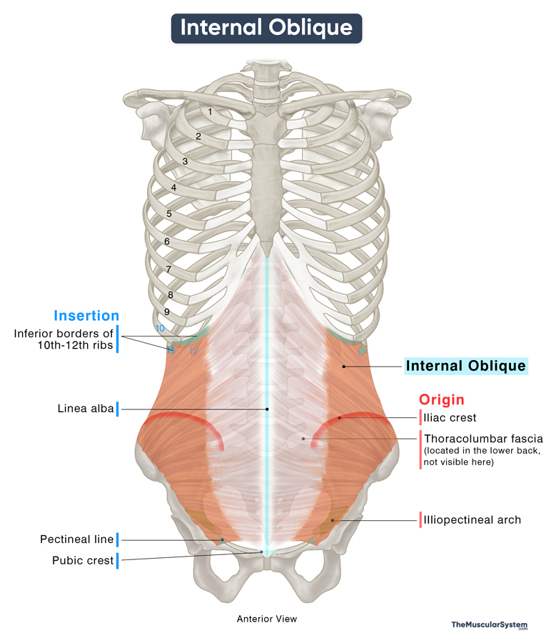
The muscle arises in three parts of muscular slips: the anterior, lateral, and posterior fibers.
Internal Oblique
Anterior Fibers
Origin: These fibers usually arise from the lateral part of the inguinal ligament. However, some newer studies suggest that they may also come from a deeper structure called the iliopectineal arch, which lies just beneath the inguinal ligament. Even though this idea is gaining attention, the inguinal ligament is still widely accepted as the main point of origin.
Insertion: The anterior fibers run horizontally and inferiorly, blending with those of the transversus abdominis, forming the conjoint tendon (also called the inguinal aponeurotic falx). This tendon inserts into the pubic crest and the pectineal line of the pubis, reinforcing the posterior wall of the inguinal canal.
Lateral Fibers
Origin: The anterior two-thirds of the iliac crest serves as the origin of the lateral fibers.
Insertion: These fibers run obliquely upward and medially, inserting into the linea alba, a fibrous band that extends along the midline of the abdomen.
Posterior Fibers
Origin: The posterior portion of the internal oblique arises from the thoracolumbar fascia, a dense connective tissue structure that separates the posterior abdominal muscles from the deep back muscles.
Insertion: The fibers ascend obliquely to attach to the 10th to 12th ribs and their costal cartilages, blending with the internal intercostal muscles.
Relations With Surrounding Muscles and Structures
The external oblique lies superficial, the transversus abdominis lies deep, and the rectus abdominis lies medial to the internal oblique. As the name suggests, most fibers of the internal oblique run obliquely, positioned perpendicular to those of the external oblique. Because it is broader, the external oblique nearly completely covers the internal oblique.
Its fleshy part contributes to the lateral abdominal wall, while its aponeurosis helps form the rectus sheath, a connective tissue structure that encloses the rectus abdominis muscle.
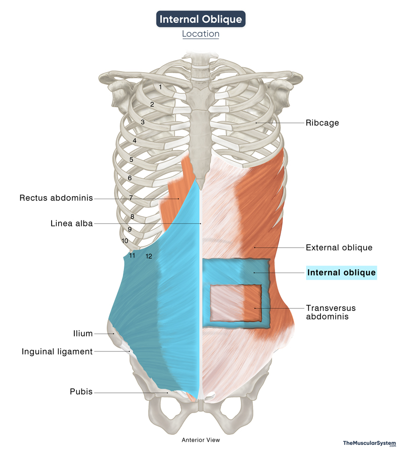
In the upper abdomen, the aponeurosis of the internal oblique splits into two layers. One layer moves forward, joining the external oblique’s aponeurosis to form the anterior portion of the rectus sheath. The other layer moves backward, merging with the transversus abdominis aponeurosis to form the posterior portion.
This arrangement changes below the arcuate line, a transition point located midway between the umbilicus and the pubic symphysis. The internal oblique’s aponeurosis no longer splits but shifts entirely to the front, reinforcing only the anterior rectus sheath. Consequently, the lower portion of the rectus abdominis is covered posteriorly only by the transversalis fascia and peritoneum rather than a true posterior rectus sheath.
The anterior fibers of the muscle contribute to the cremaster muscle in males, which surrounds the spermatic cord. In females, these fibers are often associated with the round ligament of the uterus.
Function
| Action | Flexing and rotating the trunk Compressing the abdomen to help with bodily functions |
Flexing the trunk on the side (ipsilaterally): When the muscle contracts on one side (unilaterally), it causes the trunk or torso to bend toward that side. So when the right internal oblique muscle contracts, the trunk bends to the right and vice versa. So, this muscle is active when you bend to one side to pick up your bag without bending your knees.
Flexing the trunk forward: When the muscle contracts simultaneously on both sides (bilaterally), they work with the other abdominal muscles to bend the trunk forward, like when making a formal bow.
Rotating the trunk to the sides (ipsilaterally): When the muscle contracts on one side (unilaterally), it works with the contracting external oblique muscle on the opposite side to make the torso rotate on the same side. It means that when the right internal oblique contracts, it works with the left external oblique to rotate the torso to the right. This action classifies the muscle as an ipsilateral or same-side rotator.
Role in intra-abdominal compression: Like the other muscles in the anterior-lateral abdomen, the internal oblique helps compress the organs and structures in the abdomen, helping in increasing the intra-abdominal pressure. It helps with several bodily functions like forced exhalation, defecation, and labor.
Providing structural support: Another function of the muscle is to reinforce the abdominal wall and maintain its structure so all the organs and structures stay in place. It also helps with core stability.
Antagonists
The internal oblique is an antagonist to the erector spinae muscles, specifically the iliocostalis, longissimus, and spinalis, responsible for extending the spine and torso.
It also works opposite to the diaphragm during forced exhalation. While the diaphragm pulls air into the lungs during inhalation, the internal oblique helps push air out by compressing the abdomen and pushing the diaphragm upward.
Innervation
| Nerve | Thoracoabdominal nerves (T7-T11), subcostal nerve (T12), ilioinguinal nerve (L1), and iliohypogastric nerve (L1) |
The primary innervation of the muscle comes from the lower 6 intercostal nerves, which are the thoracoabdominal nerves arising from the anterior rami of the 7th to 11th thoracic nerves, and the subcostal nerve arising from the 12th thoracic nerve.
The ilioinguinal and Ilioinguinal nerves arising from the anterior rami of the 1st lumbar spinal nerve also innervate the muscle.
Blood Supply
| Artery | Posterior intercostal and subcostal arteries |
The muscle receives its blood supply from the posterior intercostal arteries of the lower ribs and the subcostal artery arising from the 12th rib.
Additional blood supply comes from the inferior and superior epigastric arteries, superficial and deep iliac circumflex arteries, and the posterior branches of lumbar arteries.
References
- Anatomy, Abdomen and Pelvis: Abdominal Wall: NCBI.NLM.NIH.gov
- Internal Abdominal Oblique Muscle: Elsevier.com
- Muscles of the Anterolateral Abdominal Wall: ScienceDirect.com
- Internal abdominal oblique muscle: Kenhub.com
- Internal Oblique: TeachMeAnatomy.info
- Internal oblique muscle: Radiopaedia.org
- Internal abdominal oblique muscle: IMAIOS.com
Della Barnes, an MS Anatomy graduate, blends medical research with accessible writing, simplifying complex anatomy for a better understanding and appreciation of human anatomy.
- Latest Posts by Della Barnes, MS Anatomy
-
Tensor Tympani
- -
Stapedius
- -
Auricularis Posterior
- All Posts


