

Rectus Capitis Lateralis
By
Della Barnes, an MS Anatomy graduate, blends medical research with accessible writing, simplifying complex anatomy for a better understanding and appreciation of human anatomy.
Last updated:
20/11/2025Della Barnes, MS Anatomy
UX/UI Designer at - AdobeDella Barnes, an MS Anatomy graduate, blends medical research with accessible writing, simplifying complex anatomy for a better understanding and appreciation of human anatomy.
What is the Rectus Capitis Lateralis
The rectus capitis lateralis is a tiny paired muscle located deep on each side of the upper neck, at the base of the skull. It is one of the prevertebral muscles in the cervical region, along with the longus capitis, longus colli, and rectus capitis anterior. It works as a weak flexor, helping with tilting the head slightly to the side.
Anatomy
Location and Attachments
| Origin | Transverse process of the atlas bone |
| Insertion | Jugular process of the occipital bone |
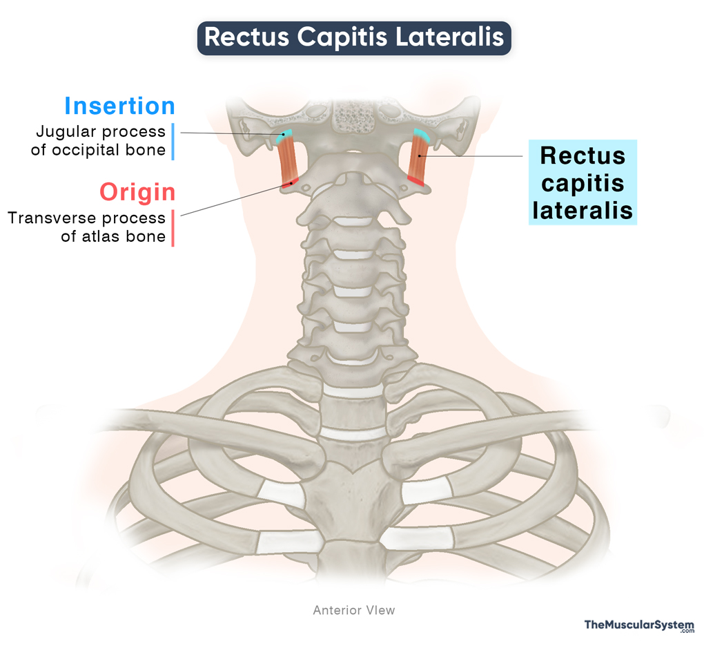
Origin
The muscle originates via narrow tendons from the superior surface of the transverse process of the first cervical vertebra, the atlas.
Insertion
From their origin, the muscle fibers ascend to form a small belly that inserts into the inferior surface of the jugular process, a small bony projection on each side at the base of the occipital bone.
Relations With Surrounding Muscles and Structures
Muscular Relations
The muscle lies lateral to both the rectus capitis anterior and the superior part of the longus capitis, both prevertebral muscles. Posteriorly, it is related to the obliquus capitis superior, and is positioned slightly above the obliquus capitis inferior, which are both suboccipital muscles. The posterior belly of the digastric, a suprahyoid muscle, lies anterior to it.
The anterior intertransversarii of the cervical region lie deep to it, while the levator scapulae, one of the extrinsic back muscles, inserts onto the inferior surface of the transverse process of the atlas. This insertion point lies just below the origin of the rectus capitis lateralis.
Neurovascular Relations
The rectus capitis lateralis serves as an important surgical landmark in the upper neck and the base of the skull because it has several vital neurovascular relations:
- The internal jugular vein runs just behind it, separated by the carotid sheath and sometimes by the accessory nerve (CN XI).
- The occipital artery typically passes between the rectus capitis lateralis and the digastric muscle.
- Medially, the vertebral artery passes along its border as the muscle exits the transverse foramen of the atlas.
- The glossopharyngeal (CN IX), vagus (CN X), and accessory nerves descend across its anterior surface after they originate from the jugular foramen.
Function
| Action | — Maintaining the stability of the head and neck at the atlanto-occipital joint — Weakly assisting in the lateral flexion of the neck |
It helps stabilize the atlanto-occipital joint by connecting the atlas (C1) to the occipital bone of the skull, contributing to the stability of the head on the neck during movement and assisting in maintaining proper head posture.
When one side of the muscle contracts (unilateral contraction), it assists the deep neck flexors by weakly flexing or bending the neck on the same side (ipsilateral flexion).
Antagonists
Being a small muscle that produces only minimal movement, the rectus capitis lateralis does not have any direct antagonists.
Innervation
| Nerve | C1-C2 spinal nerves |
The muscle is innervated by the ventral rami of the first and second cervical spinal nerves (C1-C2).
Blood Supply
| Artery | Vertebral, ascending pharyngeal, and occipital arteries |
Blood supply to the muscle comes from branches of the vertebral, ascending pharyngeal, and occipital arteries. The vertebral artery is a branch of the subclavian artery, while the other two arteries supplying this muscle originate from the external carotid artery.
References
- Rectus Capitis Lateralis Muscle: Radiopaedia.org
- Rectus Lateralis Capitis Muscle: Elsevier.com
- Rectus Capitis Lateralis Muscle: Kenhub.com
- Rectus Lateralis Capitis Muscle: IMAIOS.com
Della Barnes, an MS Anatomy graduate, blends medical research with accessible writing, simplifying complex anatomy for a better understanding and appreciation of human anatomy.
- Latest Posts by Della Barnes, MS Anatomy
-
Tensor Tympani
- -
Stapedius
- -
Auricularis Posterior
- All Posts


