

Omohyoid
By
Della Barnes, an MS Anatomy graduate, blends medical research with accessible writing, simplifying complex anatomy for a better understanding and appreciation of human anatomy.
Last updated:
15/12/2025Della Barnes, MS Anatomy
UX/UI Designer at - AdobeDella Barnes, an MS Anatomy graduate, blends medical research with accessible writing, simplifying complex anatomy for a better understanding and appreciation of human anatomy.
What is the Omohyoid
The omohyoid is a long, paired, strap-like muscle located in front of the neck, composed of two bellies connected by an intermediate tendon. It is one of the four infrahyoid muscles, along with the sternohyoid, sternothyroid, and thyrohyoid. As an extrinsic laryngeal muscle, it contributes to the hyoid bone’s movements, playing an important role in breathing and swallowing.
Anatomy
The muscle has a structure similar to that of the digastric from the suprahyoid group, with the two bellies originating from different points and joined by an intermediate tendon.
Location and Attachments
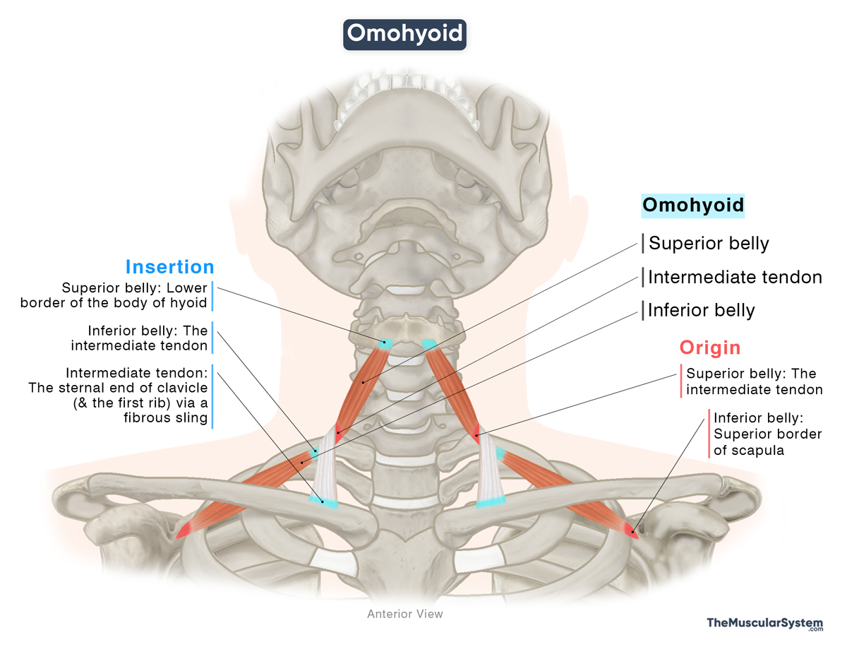
| Origin | Inferior belly: Superior border of the scapula Superior belly: The intermediate tendon |
| Insertion | Inferior belly: The intermediate tendon, which attaches to the clavicle and first rib by a fibrous sling Superior belly: Lower border of the body of the hyoid bone |
Inferior Belly
Origin
The inferior belly originates from the superior border of the scapula, on the medial side of the suprascapular notch. The point of origin often extends to the adjacent transverse scapular ligament.
Insertion
From their point of origin, the muscle fibers ascend at an oblique angle to the front. As they reach the lower part of the neck, they blend with the intermediate tendon at the level of the cricoid arch at the front of the cricoid cartilage.
The intermediate tendon itself inserts into the sternal end of the clavicle and the first rib by a fibrous fascial sling or loop.
Superior Belly
Origin
The superior belly originates from the intermediate tendon, near the internal jugular vein.
Insertion
The muscle fibers course vertically upward along the lateral side of the sternocleidomastoid muscle, and finally insert into the lower border of he body of the hyoid bone.
Relations With Surrounding Muscles and Structures
Muscular relations
Its inferior belly runs deep to the sternocleidomastoid muscle, while the superior belly courses in proximity to the sternohyoid muscle, inserting at a point lateral to the sternohyoid’s insertion. Variations may be present in some individuals in which the two muscles blend as they approach the point of insertion at the back of the hyoid bone.
Contribution to the triangles of the neck
The muscle is related to both the anterior and posterior triangles of the neck.
Anterior Triangle: The superior belly of the omohyoid contributes to two of the four divisions of the anterior triangle:
- The carotid triangle: Forms its lower front border
- The muscular triangle: Forms its upper border
These triangles contain vital nerves and arteries, including the external and internal carotid arteries (only within the carotid triangle), the internal jugular vein, and branches of the cervical plexus.
Posterior Triangle: The inferior belly divides it into two parts:
- The occipital triangle: Forms its lower border
- The subclavian triangle: Forms its upper border
Both triangles contain several important neurovascular structures, including branches of the cranial nerve XI, the cervical and brachial plexuses, the subclavian and transverse cervical arteries, part of the external jugular vein, and lymph nodes.
Relations of the intermediate tendon
The deep cervical fascia covers the intermediate tendon of the omohyoid muscle and connects the muscle to the carotid sheath. The intermediate tendon acts as a sling, connecting the two bellies to the clavicle and first rib. It also lies superficial to the internal jugular vein, making it an important surgical landmark for this vein.
Function
| Action | Depressing the hyoid bone and larynx to help with breathing |
It works with the other infrahyoid muscles to pull the hyoid bone downward. By lowering the hyoid, and with it the larynx, it helps return these structures to their resting position after swallowing. This reopens the airway to resume normal breathing.
In addition to this main role, the omohyoid is thought to assist in two supportive functions:
Because the muscle attaches to the carotid sheath, its contraction gently tugs on the sheath, increasing pressure on the internal jugular vein inside it. This action is believed to help in better drainage of blood from the head.
Similarly, contraction of the muscle also helps tighten the deep cervical fascia of the neck, preventing any soft tissue from being sucked into the airways during deep breaths.
Antagonists
As part of the infrahyoid muscle group, its action of depressing the hyoid bone is antagonized by the suprahyoid muscles, the mylohyoid, stylohyoid, digastric, and geniohyoid, which elevate the hyoid.
Innervation
| Nerve | Ansa cervicalis (C1-C3) |
Its innervation comes from the ansa cervicalis, a nerve loop that comes from the anterior rami of the first to third cervical spinal nerves (C1-C3). The inferior belly receives innervation directly from the ansa cervicalis, while the superior belly is innervated by the superior root of the ansa cervicalis, which carries fibers from the first spinal nerve (C1).
Blood Supply
| Artery | Infrahyoid branches of the superior thyroid artery |
The primary blood supply to the muscle comes from the infrahyoid branches of the superior thyroid artery, which branches from the external carotid artery. Additionally, the inferior thyroid artery, a branch of the thyrocervical trunk, also provides blood supply.
References
- Omohyoid: TeachMeAnatomy.info
- Omohyoid Muscle – Kenhub.com
- Omohyoid Muscle (Left): Elsevier.com
- Omohyoid Muscle | Function, Origin & Innervation: Study.com
- Omohyoid Muscle: Radiopaedia.org
- Omohyoid: HealthLine.com
Della Barnes, an MS Anatomy graduate, blends medical research with accessible writing, simplifying complex anatomy for a better understanding and appreciation of human anatomy.
- Latest Posts by Della Barnes, MS Anatomy
-
Tensor Tympani
- -
Stapedius
- -
Auricularis Posterior
- All Posts


