

Splenius Capitis
By
Della Barnes, an MS Anatomy graduate, blends medical research with accessible writing, simplifying complex anatomy for a better understanding and appreciation of human anatomy.
Last updated:
30/01/2026Della Barnes, MS Anatomy
UX/UI Designer at - AdobeDella Barnes, an MS Anatomy graduate, blends medical research with accessible writing, simplifying complex anatomy for a better understanding and appreciation of human anatomy.
What is the Splenius Capitis
Splenius capitis is a thick, paired band of muscle at the back of the neck, connecting the skull and the top of the vertebral column. It belongs to the group of intrinsic back muscles and is one of the primary movers of the head and neck. It belongs to the spinotransversales group, along with the splenius cervicis.
The name “splenius capitis” is derived from “splēníon,” the Greek word meaning “bandage,” and “caput,“ the Latin word for head. It refers to how the muscle wraps around the back of the neck like a bandage.
Anatomy
Location and Attachments
| Origin | Spinous processes of the C7 to T3 vertebra and the nuchal ligament |
| Insertion | The occipital bone and the mastoid process of the temporal bone |
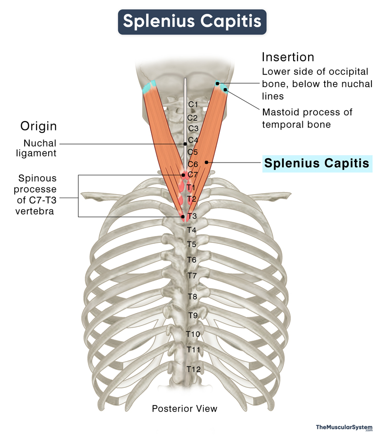
Origin
The muscle’s origin can be divided into two regions. The first region arises from the nuchal ligament at the level of the 4th to 6th cervical vertebrae (C4 to C6). This ligament is a band-like structure connecting the base of the skull to the cervical vertebrae. The muscle also originates from the spinous processes of the 7th cervical vertebra (C7) and the upper three thoracic vertebrae (T1 to T3).
Insertion
From the points of origin, the muscle fibers run obliquely upward and laterally toward the base of the skull to insert into the temporal and occipital bones. On the temporal bone, it inserts into the mastoid process, which is a bony prominence located just behind the ear. The part of the muscle that inserts into the occipital bone courses to the back of the head and inserts just below the nuchal lines on the lateral side.
Relations With Surrounding Muscles and Structures
Though part of the intrinsic or deep back muscles, splenius capitis is one of the most superficial muscles in the group. The capitis parts of two other intrinsic muscles, the semispinalis, and the longissimus muscles, lie deep to this muscle.
The trapezius, a spine muscle extending from the base of the neck to the mid-back region, is superficial to the lower part of the splenius capitis. The upper part of the muscle, which is attached to the mastoid process, lies beneath the sternocleidomastoid (SCM), one of the largest neck muscles.
Splenius capitis is one of the muscles that form the floor of the posterior triangle of the neck — an important anatomical landmark.
Function
| Action | Extending, laterally flexing, and rotating the head and neck |
The muscle supports the head and neck. When both sides (bilateral contraction) contract, it helps extend or straighten the head, such as raising the head from a lowered position or tilting it back, as you do when looking up.
When the muscle contracts on one side (unilateral contraction), it causes lateral flexion and ipsilateral rotation of the cervical spine. These movements allow you to bend your head to the side or rotate it toward the same side as the contracting muscle. For example, when you turn or tilt your head to the right, the right splenius capitis muscle is involved, and the left muscle is engaged when you turn to the left.
Antagonists
The sternocleidomastoid can be considered antagonistic to the splenius capitis. The SCM primarily functions to flex the cervical spine and assists in bowing the head forward. Additionally, when the SCM contracts on one side, it rotates the head to the opposite side, contrasting with the actions of the splenius capitis, which extends and rotates the head to the same side.
Innervation
| Nerve | Posterior rami of C2 and C3 spinal nerves |
The Splenius Capitis muscle receives innervation from the posterior or dorsal rami of the 2nd and 3rd cervical spinal nerves (C2-C3). Additionally, the 4th cervical nerve (C4) can also contribute to its innervation sometimes.
Blood Supply
| Artery | Muscular branches of the occipital artery |
The occipital artery, which branches from the external carotid artery, is responsible for the blood supply. The primary vasculature comes from the muscular branches of the occipital artery. Additionally, the deep cervical artery, which branches from the costocervical trunk of the subclavian artery, provides further support.
References
- Splenius Capitis Muscle | Origin, Insertion & Action: Study.com
- Splenius Capitis: Meddean.LUC.edu
- Splenius Capitis Muscle: GetBodySmart.com
- Splenius Capitis Muscle: Kenhub.com
- Splenius Capitis: TeachMeanAtomy.info
- Splenius Capitis Muscle: Elsevier.com
Della Barnes, an MS Anatomy graduate, blends medical research with accessible writing, simplifying complex anatomy for a better understanding and appreciation of human anatomy.
- Latest Posts by Della Barnes, MS Anatomy
-
Tensor Tympani
- -
Stapedius
- -
Auricularis Posterior
- All Posts


