

External Oblique
By
Della Barnes, an MS Anatomy graduate, blends medical research with accessible writing, simplifying complex anatomy for a better understanding and appreciation of human anatomy.
Last updated:
23/05/2025Della Barnes, MS Anatomy
UX/UI Designer at - AdobeDella Barnes, an MS Anatomy graduate, blends medical research with accessible writing, simplifying complex anatomy for a better understanding and appreciation of human anatomy.
What Is the External Oblique
The external oblique is a flat, broad, paired muscle located on the two sides of the human abdomen, extending from the ribcage down to the pelvis. It is the largest and the most laterally located of the three muscles in the anterior-lateral abdominal wall, with the other two being the rectus abdominis and internal oblique.
The muscle plays a vital role in the flexibility of the spinal column, helping with movements like rotation and bending of the trunk.
Anatomy
Location and Attachments
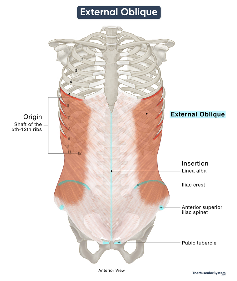
| Origin | Shaft of the 5th-12th ribs |
| Insertion | Iliac crest, anterior superior iliac spine, pubic tubercle, and linea alba |
Origin
The muscle originates as eight fleshy slips from the lower surfaces and lateral borders of the shaft of the lower eight ribs (5th-12th).
Insertion
From their point of origin, the muscle fibers run obliquely towards the midline of the body, covering the sides of the abdomen. As they travel toward the point of insertion, the muscle fibers gradually turn more aponeurotic. The change starts above the umbilical (bellybutton), near the midclavicular line, an imaginary reference line at the front of the chest wall, passing through the midpoint of the collarbone.
The upper part of the muscle inserts along the length of the linea alba – the thick aponeurotic band running down the midline of the abdominal wall. The lower part inserts into the front of the iliac crest and the anterior superior iliac spine (ASIS). The lowest fibers attach to the pubic crest and tubercle.
Relations With Surrounding Muscles and Structures
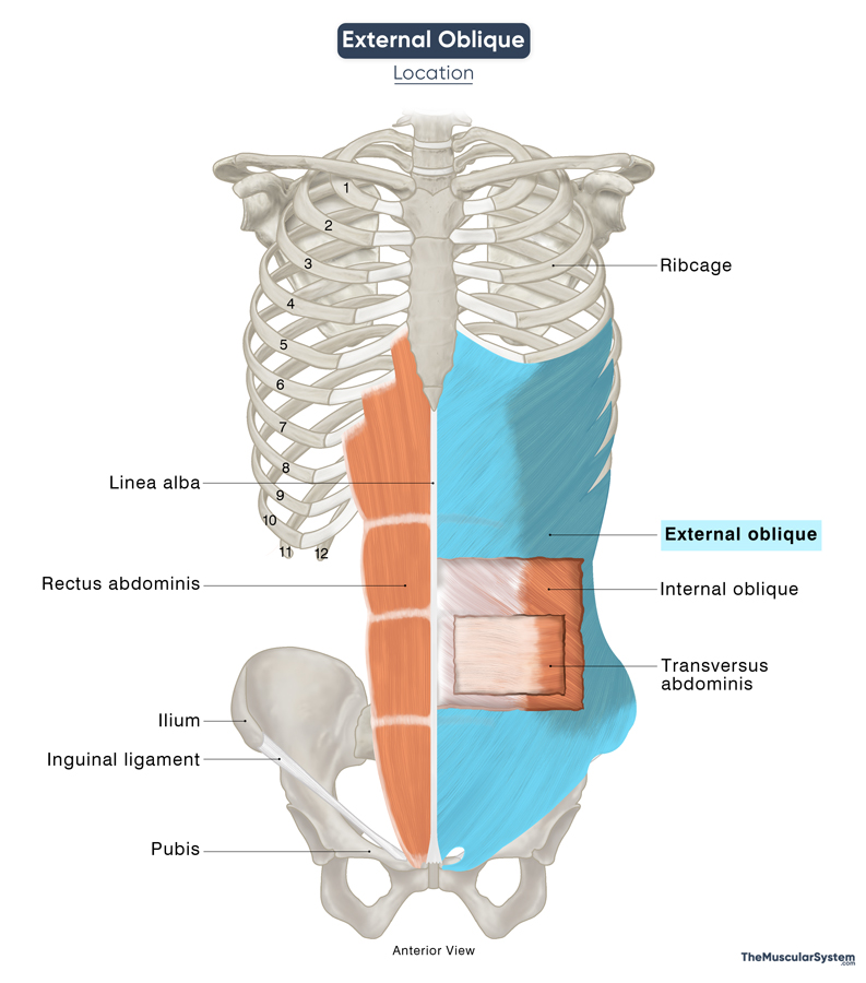
The external oblique is the most superficial of the abdominal muscles. It covers part of the lower ribcage and overlies the external intercostal muscles in that region. The internal oblique muscle lies underneath it.
As one of the largest abdominal muscles, with a broad aponeurotic portion, the external oblique contributes to the formation of several structures in the human abdomen. Its muscular part forms the lateral wall of the abdomen, while the aponeurotic portion, particularly in its upper half, contributes to the anterior layer of the rectus sheath, which encloses the rectus abdominis muscle medially.
The inferior part of the external oblique’s aponeurosis thickens and rolls inward at its lower border, forming the inguinal ligament. This fibrous band extends from the anterior superior iliac spine (ASIS) to the pubic tubercle, marking the transition between the pelvis and the thigh.
The external oblique’s aponeurosis also contributes to the anterior wall of the inguinal canal, a passage in the lower abdomen that allows the spermatic cord (in males) and the round ligament (in females) to pass from the abdomen to the external genitalia. These structures exit through the superficial inguinal ring, a V-shaped opening in the aponeurosis located above the pubic tubercle.
Posteriorly, the fleshy posterior border of the external oblique helps form the anterior boundary of the lumbar (Petit’s) triangle, an anatomical landmark in the lower back.
Function
| Action | Flexing and rotating the trunk Stabilizing the core |
1. Flexing the trunk to the same side (ipsilaterally): When the muscle contracts on one side (unilaterally), it pulls the ribcage downward, causing the trunk to bend toward the same side. So, bending your body toward the right requires the right external oblique to be active, and when you bend to the left, the left external oblique is active.
An example of this action would be when you bend to one side to pick something up from the floor without bending your knees.
2. Flexing the trunk forward: When both sides of the external oblique contract together (bilaterally), they pull the trunk forward, assisting the torso in bending forward. This muscle works with the rectus abdominis to flex the trunk.
For example, this muscle helps when you make a formal bow.
3. Rotating the trunk to the opposite side (contralaterally): The muscle contracts unilaterally and in synergy with the internal oblique on the opposite side to help rotate the trunk contralaterally. So, when the right external oblique contracts, the torso twists to the left and vice versa.
For example, when you twist your body to the right when doing a bicycle crunch, your left external oblique muscle contracts.
4. Intra-abdominal compression: Its contraction, along with other muscles in the area, helps compress the contents of the abdomen (abdominal viscera), increasing intra-abdominal pressure. This assists in physiological processes like forced exhalation, defecation, and labor.
5. Core stability and structural support: The muscle reinforces the abdominal wall, providing structural support to the abdominal organs, structures, and the trunk as a whole.
Antagonists
The muscles in the erector spinae group — iliocostalis, longissimus, and spinalis — are considered antagonistic to the external oblique because they extend the spine, counteracting its flexion.
Innervation
| Nerve | Thoraco abdominal nerves (T7-T11), subcostal nerve (T12) |
The superior part of the muscle is innervated by the thoracoabdominal nerves, arising from the anterior rami of the T7 to T11 spinal nerves. The inferior part receives innervation from the T12 thoracic nerve, also known as the subcostal nerve.
Its sensory innervation comes from the iliohypogastric nerve (L1), which is a branch of the lumbar plexus.
Blood Supply
| Artery | Lower posterior intercostal arteries, subcostal artery, deep circumflex iliac artery, superior and inferior epigastric arteries |
The superior two-thirds of the muscle receives blood supply from the lower posterior intercostal (T9-T11) and subcostal (T12) arteries. Blood supply to the inferior part comes from the deep circumflex iliac artery.
Additionally, blood supply comes from the superior and inferior epigastric arteries.
References
- External Oblique: TeachMeAnatomy.info
- External Abdominal Oblique Muscle | Anatomy & Functions: Study.com
- Abdominal Muscles: ClevelandClinic.org
- Anatomy, Abdomen and Pelvis: Anterolateral Abdominal Wall: NCBI.NLM.NIH.gov
- External Abdominal Oblique Muscle: Kenhub.com
- External Abdominal Oblique Muscle: Elsevier.com
- External Abdominal Oblique Muscle: GetBodySmart.com
Della Barnes, an MS Anatomy graduate, blends medical research with accessible writing, simplifying complex anatomy for a better understanding and appreciation of human anatomy.
- Latest Posts by Della Barnes, MS Anatomy
-
Tensor Tympani
- -
Stapedius
- -
Auricularis Posterior
- All Posts


