

Mylohyoid
By
Della Barnes, an MS Anatomy graduate, blends medical research with accessible writing, simplifying complex anatomy for a better understanding and appreciation of human anatomy.
Last updated:
04/12/2025Della Barnes, MS Anatomy
UX/UI Designer at - AdobeDella Barnes, an MS Anatomy graduate, blends medical research with accessible writing, simplifying complex anatomy for a better understanding and appreciation of human anatomy.
What is the Mylohyoid
The mylohyoid, sometimes referred to as the oral diaphragm or diaphragma oris, is a paired muscle of the neck, contributing to the floor of the oral cavity. It belongs to the group of suprahyoid muscles along with the stylohyoid, digastric, and geniohyoid. It attaches the hyoid bone to the mandible, assisting in vital actions like speaking and swallowing.
Anatomy
Location and Attachments
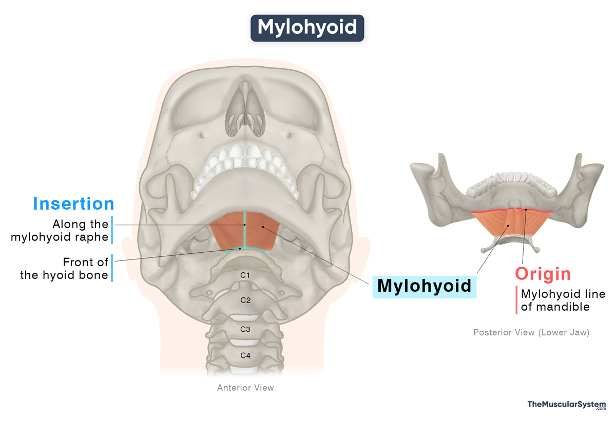
| Origin | Mylohyoid line of the mandible |
| Insertion | The front of the hyoid bone and the mylohyoid raphe |
Origin
The muscle has a broad origin from the mylohyoid line, the bony ridge on the inner surface of the mandible, on both sides. The point of origin lies just behind the molar tooth, which is where the name “mylohyoid” is derived from.
Insertion
From their origin, the muscle fibers descend medially as they form a broad muscle belly. The posterior part of the muscle inserts into the front of the hyoid bone. The medial and anterior portions attach to the mylohyoid raphe, a fibrous band extending from the mandible’s midline (symphysis menti) to the upper surface of the body of the hyoid bone.
Variations
In some cases, the mylohyoid raphe may be absent altogether. In such cases, the two mylohyoid muscles may blend with the digastric muscle’s anterior belly or with each other. Sometimes, the muscle may be absent altogether, when its role is taken up by the anterior belly of the digastric.
Relations With Surrounding Muscles and Structures
Together, the left and right mylohyoid form the boundary between the mouth above and the neck below. This earns the muscle the name oral diaphragm. Spanning between the mandible and the hyoid, the muscle forms the floor of the submental triangle, the division of the anterior triangle of the neck, just below the chin.
Superiorly, the muscle is related to the structures of the oral cavity, lying immediately deep to the geniohyoid, hyoglossus, and styloglossus muscles. The hypoglossal and lingual nerves, the lingual vessels, the submandibular ganglion, and the sublingual gland also lie superficially.
Inferior relations of the muscle include the platysma and the anterior belly of the digastric muscle. It also has the mylohyoid nerve and artery, and the facial and submenta vessels lying deep to it.
The submandibular gland curves around the posterior border of the mylohyoid, which divides the gland into the superficial and deep lobes.
Function
| Action | Elevating the floor of the oral cavity and the hyoid bone to help with actions like swallowing, speaking, and chewing |
As it forms the floor of the oral cavity and connects the mandible and the hyoid bones, the muscle plays a key role in oral and pharyngeal movements.
When the muscle contracts with the mandible staying fixed, it elevates the hyoid bone and the floor of the mouth. This helps raise the tongue and propel the food toward the pharynx during swallowing. This action of raising the tongue also helps with articulation in speech.
When the hyoid bone is fixed, this muscle works with other suprahyoid muscles, like the digastric and geniohyoid, to depress the mandible. This helps release the jaws and teeth when they are clenched together by food, helping with chewing or mastication.
Antagonists
When the mylohyoid elevates the hyoid, its movement is opposed by the infrahyoid muscles, the sternohyoid, omohyoid, sternothyroid, and thyrohyoid, which act to depress or stabilize the hyoid, also helping with swallowing and speaking.
In its action to depress the mandible, its antagonists include the primary jaw-elevating muscles, the masseter, temporalis, and medial pterygoid.
Innervation
| Nerve | Mylohyoid nerve |
The mylohyoid nerve, also known as the nerve to mylohyoid, arises from the inferior alveolar nerve and innervates this muscle. The inferior alveolar nerve itself is a branch of the mandibular nerve.
Blood Supply
| Artery | Inferior alveolar, submental, and sublingual arteries |
Blood supply to this muscle comes from the inferior alveolar artery, which is a branch of the maxillary artery. The submental artery, branching off the facial artery, and the sublingual artery, a branch of the lingual artery also provides blood supply.
References
- Mylohyoid Muscle: Kenhub.com
- Anatomy, Head and Neck, Mylohyoid Muscle: NCBI.NLM.NIH.gov
- Mylohyoid: TeachMeAnatomy.info
- Mylohyoid Muscle: Elsevier.com
- Mylohyoid Muscle: Radiopaedia.org
Della Barnes, an MS Anatomy graduate, blends medical research with accessible writing, simplifying complex anatomy for a better understanding and appreciation of human anatomy.
- Latest Posts by Della Barnes, MS Anatomy
-
Tensor Tympani
- -
Stapedius
- -
Auricularis Posterior
- All Posts


