

Coccygeus
By
Della Barnes, an MS Anatomy graduate, blends medical research with accessible writing, simplifying complex anatomy for a better understanding and appreciation of human anatomy.
Last updated:
29/05/2025Della Barnes, MS Anatomy
UX/UI Designer at - AdobeDella Barnes, an MS Anatomy graduate, blends medical research with accessible writing, simplifying complex anatomy for a better understanding and appreciation of human anatomy.
What is the Coccygeus
Coccygeus, also called ischiococcygeus, is a small, triangular, paired muscle in the pelvic region. It contributes to the pelvic floor or pelvic diaphragm alongside the levator ani muscle group. Functions of the muscle include supporting the pelvic organs and helping with coccygeal movements.
Anatomy
Location and Attachments
| Origin | Ischial spine |
| Insertion | Sacrum and coccyx |
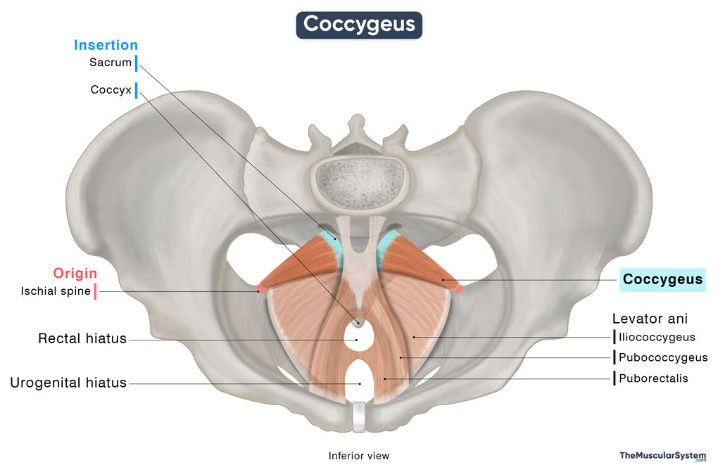
Origin
The apex of the triangular muscle marks its origin, arising from the ischial spine — the pointy, bony projection at the back of the ischium bone.
Insertion
Originating at the ischial spine, the muscle fibers of the coccygeus fan out and course medially and posteriorly, forming a triangular shape, with the base marking its point of insertion. The muscle inserts onto the lateral margins of the lower sacrum and coccyx (tailbone), typically at the level of the fourth and fifth sacral segments (S4-S5)
Relations to Other Muscular Structures
The coccygeus muscle forms the upper posterior part of the pelvic floor. It lies posterior and slightly lateral to the levator ani, particularly the iliococcygeus portion, which is the posterior-most part of the levator ani.
Superior to the coccygeus lies the piriformis, a muscle of the gluteal region. These two muscles are related near the lower border of the piriformis, and their positioning helps define part of the greater sciatic foramen. It is a bony opening that allows several important nerves and arteries to pass from the pelvis to the gluteal region, including the inferior gluteal artery, internal pudendal artery, and pudendal nerve.
On its gluteal surface, the coccygeus becomes fibrous and contributes to the sacrospinous ligament. In some individuals, the muscle may lack fleshy fibers entirely. Instead, it may be composed entirely of tendinous tissue, making it indistinguishable from the sacrospinous ligament.
On its pelvic surface lies the coccygeus plexus, a small network of nerves that supplies the pelvic region.
Function
| Action | Supporting the pelvic viscera and flexing the coccyx |
Being an integral part of the pelvic floor, the coccygeus muscle plays a vital role in supporting the organs in the lower part of the abdomen, such as the bladder, intestines, and uterus (in women).
When this muscle contracts, it pulls the contents of the lower pelvis slightly forward and upward, while also drawing the coccyx or tailbone forward. This action helps maintain the integrity of the pelvic cavity, and prevents organ prolapse, a condition where an organ slips down through a pelvic hiatus or opening like the urethra or anus.
It also assists the levator ani muscles, pubococcygeus and puborectalis, in controlling urination and defecation, especially when the abdominal pressure is high, such as during coughing, sneezing, or lifting something heavy.
Sometimes, the pressure of childbirth (or even defecation) pushes the tailbone backward to make more space. Once the process is complete, the coccygeus contracts to bring the tailbone back in its place.
Innervation
| Nerve | Anterior rami of S4-S5 |
The Coccygeus muscle receives innervation from the anterior rami of the 4th and 5th sacral nerves (S4-S5). Sometimes, the sacral nerve S3 may lend a minor contribution.
Blood Supply
| Artery | Inferior gluteal, inferior vesical, and internal pudendal arteries |
The primary blood supply to the muscle comes from the inferior gluteal, inferior vesical, and internal pudendal arteries — all three branching from the internal iliac artery.
References:
- Coccygeus Muscle: Kenhub.com
- Coccygeus: TeachMeAnatomy.info
- Coccygeus Muscle: Radiopaedia.org
- Coccygeus Muscle: Elsevier.com
- Coccygeus Muscle: IMAIOS.com
Della Barnes, an MS Anatomy graduate, blends medical research with accessible writing, simplifying complex anatomy for a better understanding and appreciation of human anatomy.
- Latest Posts by Della Barnes, MS Anatomy
-
Tensor Tympani
- -
Stapedius
- -
Auricularis Posterior
- All Posts


