

Muscles in the Hypothenar Eminence
By
Della Barnes, an MS Anatomy graduate, blends medical research with accessible writing, simplifying complex anatomy for a better understanding and appreciation of human anatomy.
Last updated:
30/05/2023Della Barnes, MS Anatomy
UX/UI Designer at - AdobeDella Barnes, an MS Anatomy graduate, blends medical research with accessible writing, simplifying complex anatomy for a better understanding and appreciation of human anatomy.
What is the Hypothenar Eminence
The hypothenar eminence is the fleshy mound on the medial side (the side of the little finger) in the palm of the hand, just above the wrist. It is formed by a group of intrinsic hand muscles in the hand’s medial palmar compartment. These muscles are collectively called the hypothenar muscles. The hypothenar region spans the length of the carpal and metacarpal bones in the 5th digit or little finger.
Names of the Hypothenar Muscles With Anatomy and Functions
The hypothenar group includes three primary muscles: abductor digiti minimi, flexor digiti minimi brevis, and opponens digiti minimi. There is a fourth muscle, the palmaris brevis, which is often included in the hypothenar group as it also contributes to the formation and functioning of the hypothenar eminence. But the first three are the main muscles in the group.
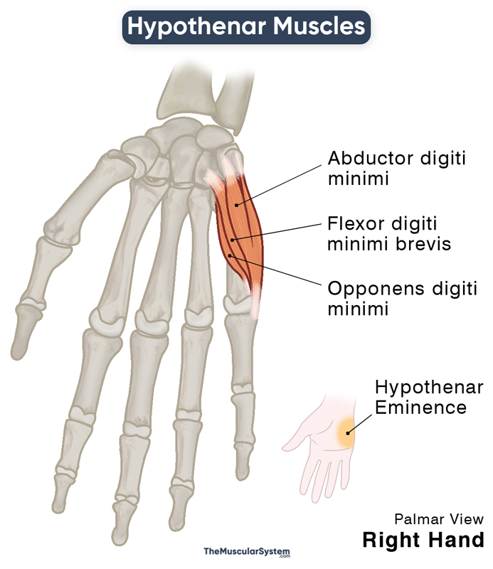
- Abductor Digiti Minimi (ADM): It is the most superficially and medially located hypothenar muscle, forming the medial or ulnar border of the hypothenar eminence. It originates from the pisiform bone, pisohamate ligament, and the adjacent flexor retinaculum. The small muscle then courses medially to insert into the base of the 5th proximal phalanx, the little finger’s proximal phalanx. This muscle works on the 5th metacarpophalangeal joint to abduct the little finger.
- Flexor Digiti Minimi Brevis (FDMB): This Muscle is located lateral to the ADM, originating from the hook of the hamate and its adjacent flexor retinaculum. It has the same insertion point as the ADM, at the base of the 5th proximal phalanx, on its medial side. Its function is to help flex the little finger.
- Opponens Digiti Minimi (ODM): The deepest hypothenar muscle, it lies deep to both the ADM and FDMB. It shares its point of origin with the FDMB, originating from the hook of the hamate and flexor retinaculum. This muscle then courses towards the 5th proximal phalanx to insert along its ulnar or medial surface. Its primary functions involve flexion and opposition of the little finger.
As mentioned above, the palmaris brevis is sometimes referred to as the 4th hypothenar muscle. It originates from the flexor retinaculum and palmar aponeurosis and inserts into the dermal layer of the skin of the palm on the ulnar side. Its function is to tighten the palmar aponeurosis to tighten the hand’s grip.
Innervation
All three hypothenar muscles are innervated by the deep branch of the ulnar nerve, which arises from the nerve roots C8 and T1.
The palmaris brevis, innervated by the ulnar nerve’s superficial branch, protects the nerve by allowing it to pass safely from underneath it as it enters the hand.
Blood Supply
The primary blood flow to the hypothenar eminence comes from the ulnar artery, specifically the superficial palmar arch. Additional blood supply comes from the deep palmar arch of the radial artery, which anastomoses with the superficial palmar arch.
References
- Anatomy, Shoulder and Upper Limb, Hand Hypothenar Eminence: NCBI.NLM.NIH.gov
- Hypothenar Muscles: KenHub.com
- Muscles of the Hand: TeachMeAnatomy.info
- Hypothenar Eminence: RadioPaedia.org
- Hypothenar Muscles: OUHSC.edu
Della Barnes, an MS Anatomy graduate, blends medical research with accessible writing, simplifying complex anatomy for a better understanding and appreciation of human anatomy.
- Latest Posts by Della Barnes, MS Anatomy
-
Tensor Tympani
- -
Stapedius
- -
Auricularis Posterior
- All Posts


