

Innermost Intercostals
By
Della Barnes, an MS Anatomy graduate, blends medical research with accessible writing, simplifying complex anatomy for a better understanding and appreciation of human anatomy.
Last updated:
11/06/2024Della Barnes, MS Anatomy
UX/UI Designer at - AdobeDella Barnes, an MS Anatomy graduate, blends medical research with accessible writing, simplifying complex anatomy for a better understanding and appreciation of human anatomy.
What Are the Innermost Intercostals
The innermost intercostals form the deepest layer of the muscles that occupy the intercostal spaces in the human rib cage. Like the other two muscles in this group, the internal and external intercostals, the innermost layer also plays an important role in respiration and maintaining the stability of the thoracic cavity.
Anatomy
Location and Attachments
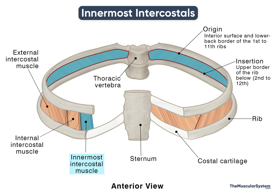
| Origin | The interior surface and infero-posterior borders of the 1st to 11th ribs |
| Insertion | The superior border of the 2nd to 12th ribs |
Origin
Each innermost intercostal muscle arises from the inner surface and the lower-back border of the costal groove of the rib immediately above it.
Insertion
From their insertion point, the muscle fibers run in a lateral downward and backward direction — parallel to the innermost intercostals and perpendicular to the external intercostals. Then, they are inserted into the upper border of the corresponding rib below.
Relations With Surrounding Muscles and Structures
The internal intercostal muscles lie immediately superior to the innermost intercostals, separated by the neurovascular bundle of each intercostal space. The external intercostals lie superior to both the innermost and internal intercostals. The parietal pleura and the endothoracic fascia lie deep to the innermost intercostals.
Unlike the muscles in the other two intercostal layers, the innermost intercostals are not all developed similarly and don’t fill the intercostal spaces equally. The muscles in the lower ribs are more developed, becoming less prominent towards the upper ribs. Usually, even the most developed muscles cover only the middle two-thirds or two-quarter of the intercostal spaces.
Function
| Action | Helping in respiration by fixing the position of the ribs |
Though considered the weakest of the three intercostals, the functioning of these muscles is similar to that of the internal intercostals. They help depress the ribcage to decrease its volume and expel air from the lungs during forced exhalation.
Additionally, these muscles maintain a constant tonic contraction. This enhances the rigidity of the chest wall, making it easy for the diaphragm to manipulate during breathing.
Innervation
| Nerve | Corresponding branches of the 1st to 11th intercostal nerves |
Like the other intercostals, the innermost layer is also innervated by the 1st to 11th intercostal nerves, originating from the thoracic spinal nerves.
Blood Supply
| Artery | Anterior and posterior intercostal arteries |
Blood supply comes from the anterior and posterior intercostal arteries, while the anterior and posterior intercostal veins do the drainage. These veins drain into the azygos and brachiocephalic veins, both of which ultimately drain into the superior vena cava.
References
- Innermost Intercostals: PrimaryCareNotebook.com
- Innermost Intercostal Muscles: Elsevier.com
- Innermost Intercostal Muscles: KenHub.com
- Innermost Intercostal Muscle: ScienceDirect.com
- Innermost Intercostals: Meddean.LUC.edu
- Innermost Intercostal Muscles: Radioaedia.org
Della Barnes, an MS Anatomy graduate, blends medical research with accessible writing, simplifying complex anatomy for a better understanding and appreciation of human anatomy.
- Latest Posts by Della Barnes, MS Anatomy
-
Tensor Tympani
- -
Stapedius
- -
Auricularis Posterior
- All Posts


