

Digastric
By
Della Barnes, an MS Anatomy graduate, blends medical research with accessible writing, simplifying complex anatomy for a better understanding and appreciation of human anatomy.
Last updated:
29/11/2025Della Barnes, MS Anatomy
UX/UI Designer at - AdobeDella Barnes, an MS Anatomy graduate, blends medical research with accessible writing, simplifying complex anatomy for a better understanding and appreciation of human anatomy.
What is the Digastric
Digastric is a paired muscle located at the front of the neck. The muscle has two prominent bellies, with its name being derived from the Greek words “dis” meaning two, and “gaster” meaning belly. It is one of the four suprahyoid muscles, along with the stylohyoid, mylohyoid, and geniohyoid. Together, they help with actions like chewing, swallowing, and speaking.
Anatomy
The muscle is divided into two bellies, the anterior and posterior bellies, joined by the intermediate tendon.
Location and Attachments
| Origin | Anterior belly: The digastric fossa of the mandible Posterior belly: The mastoid notch of the temporal bone |
| Insertion | Superior surface of the hyoid bone via the intermediate tendon |
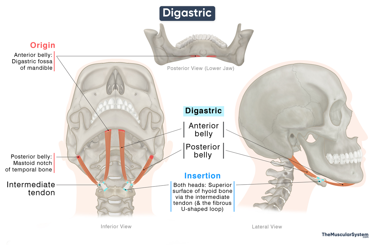
Origin
The anterior belly originates from the digastric fossa on the inner surface of the mandible, near the symphysis menti, where the two sides of the mandible bone fuse with each other.
The posterior belly originates from the medial side of the mastoid notch of the temporal bone.
Insertion
From their points of origin, the anterior belly courses posteriorly downward, and the posterior belly courses anteriorly downward to meet at the intermediate tendon, just above the hyoid bone.
The intermediate tendon inserts into the upper surface of the body and the greater cornu of the hyoid bone via a fibrous sling or a U-shaped loop of fibrous tissue that allows the tendon to glide during movement.
Relations With Surrounding Muscles and Structures
As it descends from the mastoid region, the posterior belly runs just behind the stylohyoid muscle, eventually piercing through its fibers before merging with the intermediate tendon. It also lies behind the parotid gland, which is the largest salivary gland in the human body.
Deep to the posterior belly lie several major blood vessels and nerves of the upper neck, including the carotid arteries, internal jugular vein, and the vagus, hypoglossal, and glossopharyngeal nerves.
The anterior belly lies superficial to the mylohyoid muscle.
The two bellies of the muscle divide the anterior triangle of the neck into three smaller regions:
- Submental Triangle: Lying along the midline in front of the neck, this region is bounded by the anterior bellies on each side, with the hyoid, mandible, and the mylohyoid muscle forming the other boundaries. It contains the submental lymph nodes, the submental vein, and the anterior jugular vein.
- Submandibular Triangle: Bounded by both bellies of the digastric and the lower border of the mandible, it encloses the submandibular gland and associated lymph nodes. It is also known as the digastric triangle.
- Carotid Triangle: The posterior belly forms its upper boundary, while the sternocleidomastoid and omohyoid muscles form its posterior and anterior borders. It houses key vascular structures, including the internal and external carotid arteries.
Function
| Action | Depressing the mandible to open the jaw to help with chewing, swallowing, and speech |
Because of its attachments to the mandible, temporal bone, and hyoid, the digastric muscle plays an important role in jaw and hyoid movements.
When the hyoid is stabilized by the infrahyoid muscles, the digastric, along with the other suprahyoid muscles, depresses the mandible. This action opens the jaw, which is essential for speaking, chewing, and swallowing. It can occur only when the primary jaw-closing muscles, such as the masseter and temporalis, are relaxed.
When the mandible is held in place, the digastric elevates the hyoid and indirectly raises the larynx. This elevation helps protect the airway during swallowing and supports normal breathing.
Antagonists
Muscles that close the jaw, mainly the masseter, temporalis, and medial pterygoid, are considered antagonistic to the jaw-opening action of the digastric. Additionally, the infrahyoid muscles, the sternohyoid, omohyoid, sternothyroid, and thyrohyoid, are also antagonistic to this muscle as they work to depress the hyoid bone.
Innervation
| Nerve | Anterior belly: Mylohyoid nerve (CN V₃) Posterior belly: Facial nerve (CN VII) |
The anterior and posterior bellies of the digastric muscle arise from different embryonic structures, which is why they are supplied by different cranial nerves. The anterior belly receives its innervation from the mylohyoid nerve, which is a branch of the inferior alveolar nerve from the mandibular division of the trigeminal nerve (CN V₃), while the posterior belly is innervated by the digastric branch of the facial nerve (CN VII).
Blood Supply
| Artery | Anterior belly: Submental artery Posterior belly: Occipital and posterior auricular arteries |
The two bellies also receive blood from different arterial branches of the external carotid artery. The anterior belly receives blood supply from the submental branch of the facial artery, whereas the occipital and posterior auricular arteries vascularize the posterior belly.
References
- Anatomy, Head and Neck, Digastric Muscle: NCBI.NLM.NIH.gov
- Digastric Muscle (Left): Elsevier.com
- Digastric Muscle: Kenhub.com
- Digastric Muscle: Radiopaedia.org
- Digastric Muscle Origin & Insertion: Study.com
Della Barnes, an MS Anatomy graduate, blends medical research with accessible writing, simplifying complex anatomy for a better understanding and appreciation of human anatomy.
- Latest Posts by Della Barnes, MS Anatomy
-
Tensor Tympani
- -
Stapedius
- -
Auricularis Posterior
- All Posts


