

Triceps Surae
By
Della Barnes, an MS Anatomy graduate, blends medical research with accessible writing, simplifying complex anatomy for a better understanding and appreciation of human anatomy.
Last updated:
30/10/2025Della Barnes, MS Anatomy
UX/UI Designer at - AdobeDella Barnes, an MS Anatomy graduate, blends medical research with accessible writing, simplifying complex anatomy for a better understanding and appreciation of human anatomy.
What is the Triceps Surae
The triceps surae is a large, three-headed muscle group located at the back of the lower leg, stretching from just below the knee down to the heel. It forms the prominent bulk of the calf and is often referred to as the calf muscle.
This muscle group is primarily responsible for plantar flexion at the ankle, a movement essential for walking, running, and other activities that propel the body forward.
Anatomy
Two muscles in the superficial posterior compartment of the lower leg merge to form the triceps surae:
| Muscle | Origin | Innervation | Blood Supply |
|---|---|---|---|
| Soleus | Soleal line and the middle third of the medial border of the tibiaHead and upper quarter of the posterior surface of the fibular shaft | Tibial nerve (L5-S2) | Sural, posterior tibial, and peroneal arteries |
| Gastrocnemius | Lateral head: Posterior surface of the lateral condyle of the femur Medial head: Posterior surface of the medial condyle and popliteal surface of the femur | Tibial nerve (S1-S2) | Sural artery |
Insertion of the Common Tendon
The medial and lateral heads of the gastrocnemius unite just below the knee to form a single muscle belly that shapes the upper calf. At this level, their margins also contribute to the lower medial and lateral boundaries of the popliteal fossa, the diamond-shaped space behind the knee.
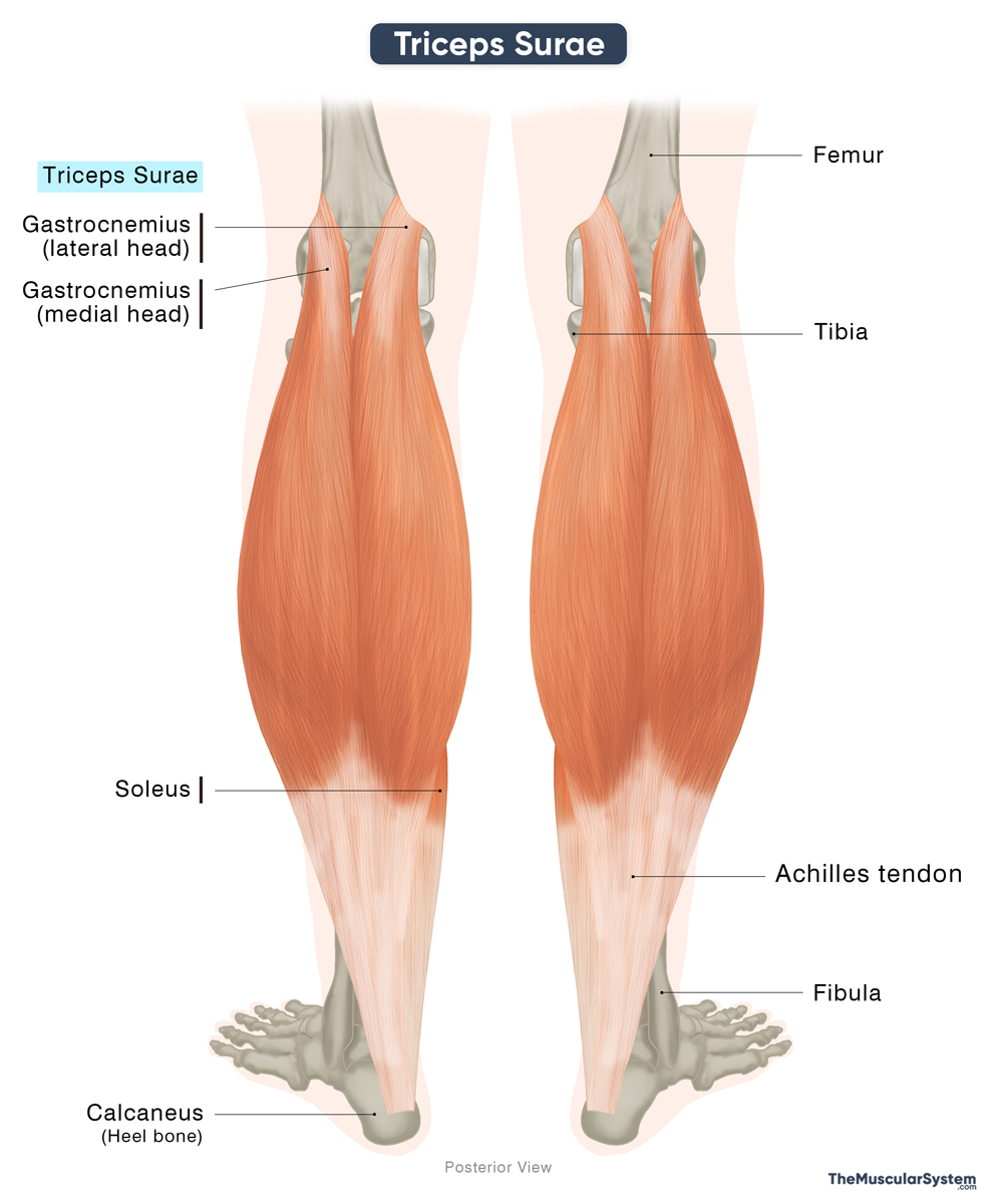
In the distal third of the lower leg, just above the ankle joint, fibers of the soleus merge with the gastrocnemius tendon. The three heads thus converge to form a common tendon, the Achilles tendon, also known as the calcaneal tendon, the thickest and strongest in the body. It then inserts onto the posterior surface of the calcaneus or heel bone.
Relations With Surrounding Muscles and Structures
Being the most superficial muscle group in the calf, the triceps surae lies deep only to the skin and the deep fascia of the leg. The plantaris muscle, also part of the superficial posterior compartment, courses between the gastrocnemius and soleus. Plantaris is not considered a component of the triceps surae. It is subject to anatomical variations and may even be absent in some individuals (around 10%). It often contributes to the Achilles tendon.
The muscles of the deep posterior compartment, the flexor digitorum longus, flexor hallucis longus, and tibialis posterior, along with the posterior tibial vessels and tibial nerve, are separated from the triceps surae by the transverse intermuscular septum.
Function
The primary action of the muscles that form the triceps surae is producing plantar flexion at the ankle, which allows the foot to push off the ground during walking, running, and jumping.
The gastrocnemius, which crosses both the knee and ankle joints, mainly contributes to powerful movements that require a lot of force, like suddenly changing direction or starting to sprint. On the other hand, the soleus, acting only at the ankle, provides endurance and stability when walking or standing.
Together, the triceps surae stabilizes the ankle, supports balance, and helps the body move efficiently by controlling shifts in weight and coordinating force during locomotion.
References
- Triceps Surae Muscle (Left): Elsevier.com
- Triceps Surae Muscle: Kenhub.com
- Triceps Surae Muscle: IMAIOS.com
- Triceps Surae Muscle: Radiopaedia.org
Della Barnes, an MS Anatomy graduate, blends medical research with accessible writing, simplifying complex anatomy for a better understanding and appreciation of human anatomy.
- Latest Posts by Della Barnes, MS Anatomy
-
Tensor Tympani
- -
Stapedius
- -
Auricularis Posterior
- All Posts


