

Fibularis Longus
By
Della Barnes, an MS Anatomy graduate, blends medical research with accessible writing, simplifying complex anatomy for a better understanding and appreciation of human anatomy.
Last updated:
10/09/2025Della Barnes, MS Anatomy
UX/UI Designer at - AdobeDella Barnes, an MS Anatomy graduate, blends medical research with accessible writing, simplifying complex anatomy for a better understanding and appreciation of human anatomy.
What is the Fibularis Longus
The fibularis longus, also known as the peroneus longus, is a long, thin muscle located on the lateral side of the lower leg. It originates just below the knee and extends down to the foot. It forms the lateral compartment of the leg along with the fibularis brevis muscle.
Its primary function is to evert the foot, while it also contributes to plantarflexion. Together, these actions provide stability and efficiency during the gait cycle.
Anatomy
Location and Attachments
| Origin | The lateral side of the head and the upper two-thirds of the fibular shaft |
| Insertion | The base of the first metatarsal and the medial cuneiform |
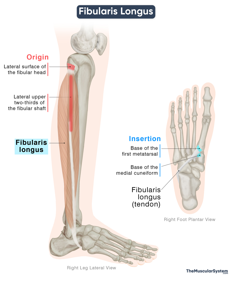
Origin
The Fibularis Longus muscle originates via tendinous attachments to multiple points:
- The head and the upper two-thirds of the lateral surface of the body of the fibula. This is the primary point of origin.
- The deep surface of the deep fascia of the leg.
- The intermuscular septa between the anterior and posterior compartments.
- Occasionally, a few fibers from the lateral condyle of the tibia.
Insertion
From its origin, the muscle runs down the outer side of the fibula as a thick, fleshy belly, which narrows back into a tendon near the ankle.
Here, the tendon takes its first turn to pass behind the lateral malleolus (the prominent bony bump at the lower end of the fibula) and underneath the superior fibular retinaculum, which holds it in place.
Next, it continues forward to the cuboid bone and makes its second turn to hook around a groove on the underside of the bone. This groove is roofed by the long plantar ligament, forming a tunnel; sometimes a fibrocartilaginous sesamoid, or even a small sesamoid bone, may develop here to reduce friction.
After this turn, the tendon runs diagonally across the sole from the outer side toward the inner side. Finally, it inserts into the base of the medial cuneiform and the first metatarsal, the bones that form the foundation of the big toe. Occasionally, a few slips extend to the second metatarsal.
Relations With Surrounding Muscles and Structures
The muscle is positioned superficially and laterally to the soleus and flexor hallucis longus of the posterior compartment, while the extensor digitorum longus of the anterior compartment lies in front of it.
On its proximal side, between its attachments on the head and shaft of the fibula, there is a small interval through which the common fibular nerve passes as it winds to the front of the leg.
At the distal side, the fibularis longus tendon meets the tendon of fibularis brevis at the lateral malleolus. From here, they share a common synovial sheath with the tendon of the fibularis brevis, as both pass beneath the superior fibular retinaculum. They are separated at the lateral aspect of the calcaneus by the fibular trochlea, with the fibularis brevis running above it, while fibularis longus courses underneath.
Function
| Action | Eversion and plantar flexion of the foot, and supporting the arches of the foot |
Eversion at the subtalar joint
It is one of the primary muscles responsible for eversion of the foot at the subtalar joint, working together with the fibularis brevis and fibularis tertius. This movement allows the sole of the foot to turn outward, away from the rest of the body. It provides lateral stability during walking and also helps the foot adapt to uneven ground.
Plantar flexion at the ankle
It contributes to plantar flexion at the ankle, together with posterior compartment muscles like gastrocnemius, soleus, and tibialis posterior. It’s the movement of the foot where you point the foot downward, like when standing on tiptoes. This movement helps during walking and running, particularly in the push-off phase.
Supporting the longitudinal and transverse arches of the foot
Since the muscle’s tendon passes diagonally across the sole, it assists in maintaining the shape of both the longitudinal and transverse arches, ensuring proper distribution of body weight through the foot during standing and movement.
Antagonists
The fibularis longus is opposed in its eversion by the tibialis anterior and tibialis posterior, both of which invert the foot. Its plantarflexion is antagonized by the tibialis anterior, which dorsiflexes the foot.
Innervation
| Nerve | Superficial fibular nerve (L5-S1) |
The muscle is mainly innervated by the superficial fibular nerve, which rises from the common fibular nerve, from the fifth lumbar and first sacral nerve roots.
Blood Supply
| Artery | Fibular artery |
The primary blood supply to the muscle is provided by the fibular artery, a branch of the tibiofibular trunk, itself a continuation of the popliteal artery.
References
- Anatomy, Bony Pelvis and Lower Limb: Calf Peroneus Longus Muscle: NCBI.NLM.NIH.gov
- Peroneus Longus: Rad.UW.edu
- Fibularis (Peroneus) Longus Muscle: Kenhub.com
- Fibularis (Peroneus) Longus: TeachMeAnatomy.info
- Fibularis Longus Muscle: Elsevier.com
- Fibularis Longus Muscle – Attachments & Action: GetBodySmart.com
Della Barnes, an MS Anatomy graduate, blends medical research with accessible writing, simplifying complex anatomy for a better understanding and appreciation of human anatomy.
- Latest Posts by Della Barnes, MS Anatomy
-
Tensor Tympani
- -
Stapedius
- -
Auricularis Posterior
- All Posts


