

Gluteus Maximus
By
Della Barnes, an MS Anatomy graduate, blends medical research with accessible writing, simplifying complex anatomy for a better understanding and appreciation of human anatomy.
Last updated:
18/08/2025Della Barnes, MS Anatomy
UX/UI Designer at - AdobeDella Barnes, an MS Anatomy graduate, blends medical research with accessible writing, simplifying complex anatomy for a better understanding and appreciation of human anatomy.
What is the Gluteus Maximus
The gluteus maximus is a large, fleshy, paired muscle that forms the main part of the human buttocks. Roughly quadrilateral in shape, it lies superficial to most other muscles and structures in the region. It is the largest muscle in the human body, serving as a primary extensor of the thigh, and forming most of the area of the buttocks we sit on.
Together with the gluteus medius and gluteus minimus, it is sometimes informally referred to as the “glutes.”
Anatomy
Location and Attachments
| Origin | — Bony attachments: Gluteal surface of the ilium between the iliac crest and posterior gluteal line, and the back and sides of the sacrum and coccyx — Aponeurotic attachments: The thoracolumbar fascia and sacrotuberous ligament |
| Insertion | The iliotibial tract and the gluteal tuberosity of the femur |
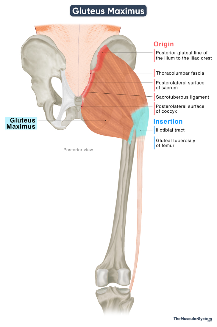
Origin
As the largest muscle in the body, the gluteus maximus has a broad origin spanning several bony and fascial landmarks. Its fibers arise from the gluteal surface of the ilium, posterior to the posterior gluteal line, and extend upward and laterally to the iliac crest. A significant portion also originates from the posterior and lateral surfaces of the sacrum and coccyx.
In addition to these bony attachments, some fibers arise from the thoracolumbar fascia and the sacrotuberous ligament. The gluteal aponeurosis (the fascia over the gluteus medius) also provides a minor point of origin.
Insertion
From its origin, the muscle fibers course obliquely downward and laterally toward the hip joint. The muscle belly can be divided into two parts based on its insertion points.
The larger, upper part of the muscle, which forms most of the buttocks, tapers into a thick, flat tendon that passes around the greater trochanter of the femur to insert into the iliotibial tract. It is an aponeurotic band that extends from the inserting fibers of the tensor fasciae latae.
The fibers from the lower part of the muscle converge into a narrower tendon that inserts on the gluteal tuberosity on the posterior femoral shaft, just above the linea aspera. This insertion lies between the insertion of the adductor magnus and the origin of the vastus lateralis. In some individuals, the third trochanter of the femur also serves as a minor point of attachment, though it is not always present.
Relations With Surrounding Muscles and Structures
Apart from being the largest, it is also the most superficially located of the three gluteal muscles. There is only a thin fascia (the superficial part of the fascia lata) separating the muscle from the subcutaneous tissue, the deepest layer of the skin.
On its anterior (deep) surface, the gluteus maximus is related to several muscles and structures, covering their posterior aspects within the gluteal region. It overlies most of the short muscles of the lateral rotator group, including the piriformis, the obturator internus, quadratus femoris, and the two gemelli muscles. It also covers the proximal portion of the gluteus medius, parts of the hamstring muscles, and the posterior aspect of the hip joint.
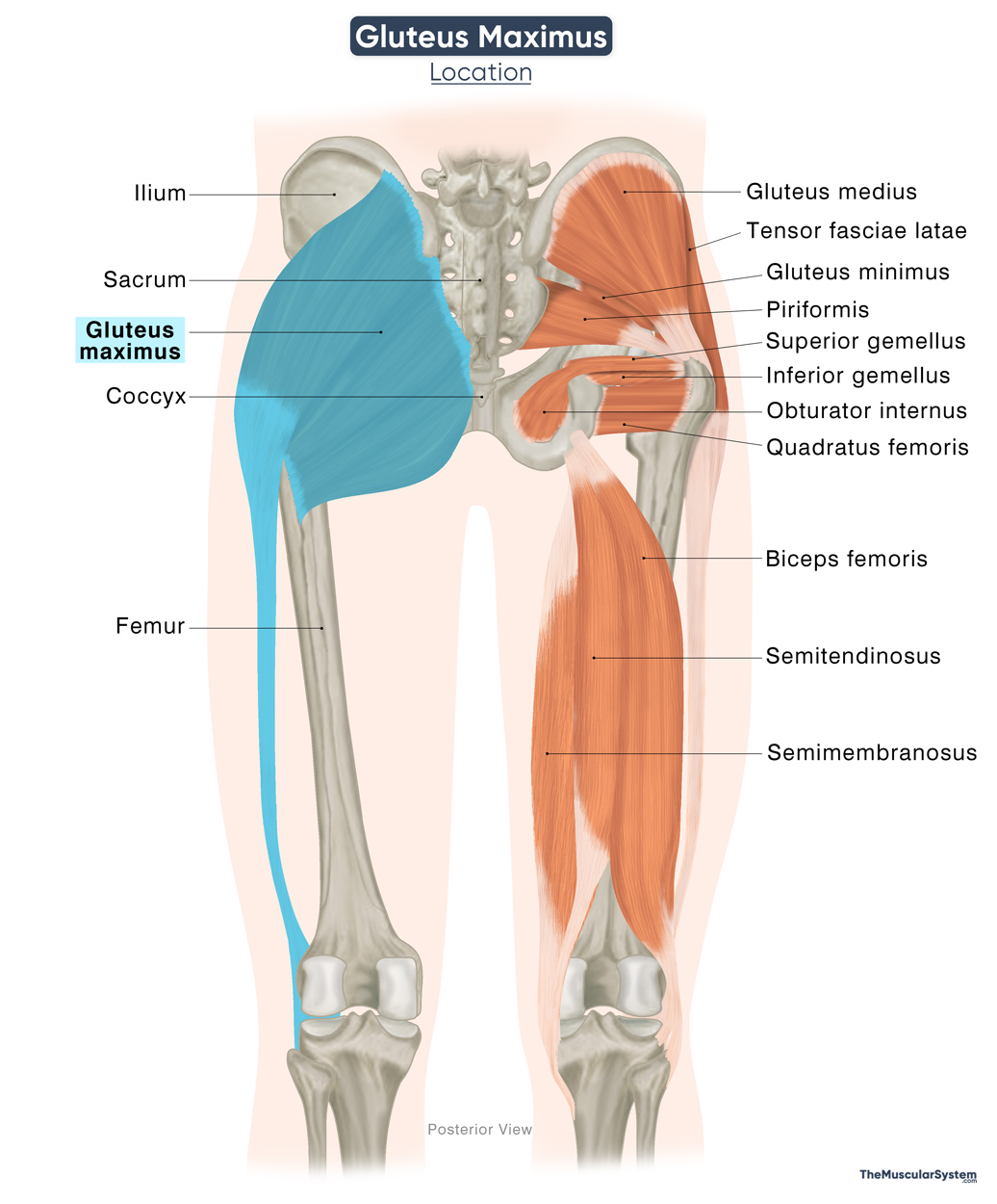
Three bursae are interposed between the muscle and the underlying bony structures to reduce friction:
- The trochanteric bursa: between the gluteus maximus and the greater trochanter.
- The ischial bursa: Between the muscle and the ischial tuberosity.
- The gluteofemoral bursa: Between the iliotibial tract and the vastus lateralis muscle. Though not directly beneath the muscle belly, it is functionally associated with the muscle.
Function
| Action | Extending, externally rotating, and abducting the thigh at the hip joint |
It is one of the most powerful muscles in the human body. Its large size, which is a distinctive feature of human musculature, along with its strength over the hip and trunk, is believed to contribute significantly to humans’ ability to stand erect. Other ape species usually have a flatter pelvis with a smaller and less powerful gluteus maximus.
The muscle primarily functions as an extensor and lateral rotator of the thigh, while also assisting other muscles in additional movements of the hip and pelvis.
1. Extending the thigh (primary function)
It stretches when the thigh moves forward (hip flexion) and contracts when the thigh moves backward (hip extension). It is a powerful hip extensor, but typically activates only when force is needed. This muscle is key in movements like rising from a chair, climbing stairs, or sprinting. During quiet standing, however, it remains mostly inactive.
It works in coordination with the hamstrings and is often referred to as an “antigravity muscle” because of its role in helping us stand upright.
2. Rotating the thigh laterally
While not a primary lateral rotator, the gluteus maximus assists muscles like the piriformis and obturators in laterally or externally rotating the thigh at the hip.
3. Abducting the thigh
The upper part of the muscle helps with the abduction of the thigh, which means moving the thigh away from the midline, like when you step sideways. The gluteus maximus also helps the medius and minimus muscles in balancing the weight of the torso on the hips when you are standing on one leg.
4. Adducting the thigh (minor role)
The lower fibers play a minor, secondary role in thigh adduction (bringing the thigh back toward the midline of the body), working alongside the primary adductors like the adductor group, pectineus, and gracilis.
5. Stabilizing the hip region
Because the gluteus maximus spans a large area, from the ilium to the hip and even to the knee via the iliotibial tract, the gluteus maximus helps stabilize the femoral head in the hip joint, and also the femur bone along the side of the thigh and knee joint.
Antagonists
Since extending the thigh at the hip is the primary function of this muscle, its primary antagonists are the hip flexors, the iliacus, and the psoas.
Innervation
| Nerve | Inferior gluteal nerve (L5-S2) |
The inferior gluteal nerve innervates this muscle. It originates from the dorsal branches of the anterior rami of the 5th lumbar (L5) and the 1st and 2nd sacral (S1–S2) nerves of the sacral plexus.
Blood Supply
| Artery | Superior and inferior gluteal arteries |
Blood supply to the gluteus maximus comes from the superior and inferior gluteal arteries. These are terminal branches of the posterior and anterior divisions of the internal iliac artery, respectively.
References
- Gluteus Maximus – Attachments, Actions & Innervation: GetBodySmart.com
- Anatomy, Bony Pelvis and Lower Limb, Gluteus Maximus Muscle: NCBI.NLM.NIH.gov
- Gluteus Maximus Muscle | Definition, Function & Structure: Study.com
- Gluteus Maximus Muscle: Kenhub.com
- Muscles of the Gluteal Region: Teachmeanatomy.info
- Gluteus Maximus Muscle: Elsevier.com
- Gluteal Muscles (Glutes): Clevelandclinic.org
Della Barnes, an MS Anatomy graduate, blends medical research with accessible writing, simplifying complex anatomy for a better understanding and appreciation of human anatomy.
- Latest Posts by Della Barnes, MS Anatomy
-
Tensor Tympani
- -
Stapedius
- -
Auricularis Posterior
- All Posts


