

Quadratus Femoris
By
Della Barnes, an MS Anatomy graduate, blends medical research with accessible writing, simplifying complex anatomy for a better understanding and appreciation of human anatomy.
Last updated:
17/07/2025Della Barnes, MS Anatomy
UX/UI Designer at - AdobeDella Barnes, an MS Anatomy graduate, blends medical research with accessible writing, simplifying complex anatomy for a better understanding and appreciation of human anatomy.
What is the Quadratus Femoris
The quadratus femoris is a small, paired muscle located deep in the gluteal region, or the buttocks. It earns its name from its rectangular or quadrilateral shape. It is the most inferiorly located muscle in the lateral hip rotators group, with other muscles in the group including the piriformis, the obturator externus and internus, and the gemelli muscles.
Anatomy
Location and Attachments
| Origin | Lateral surface of the ischial tuberosity |
| Insertion | Quadrate tubercle of the femur |
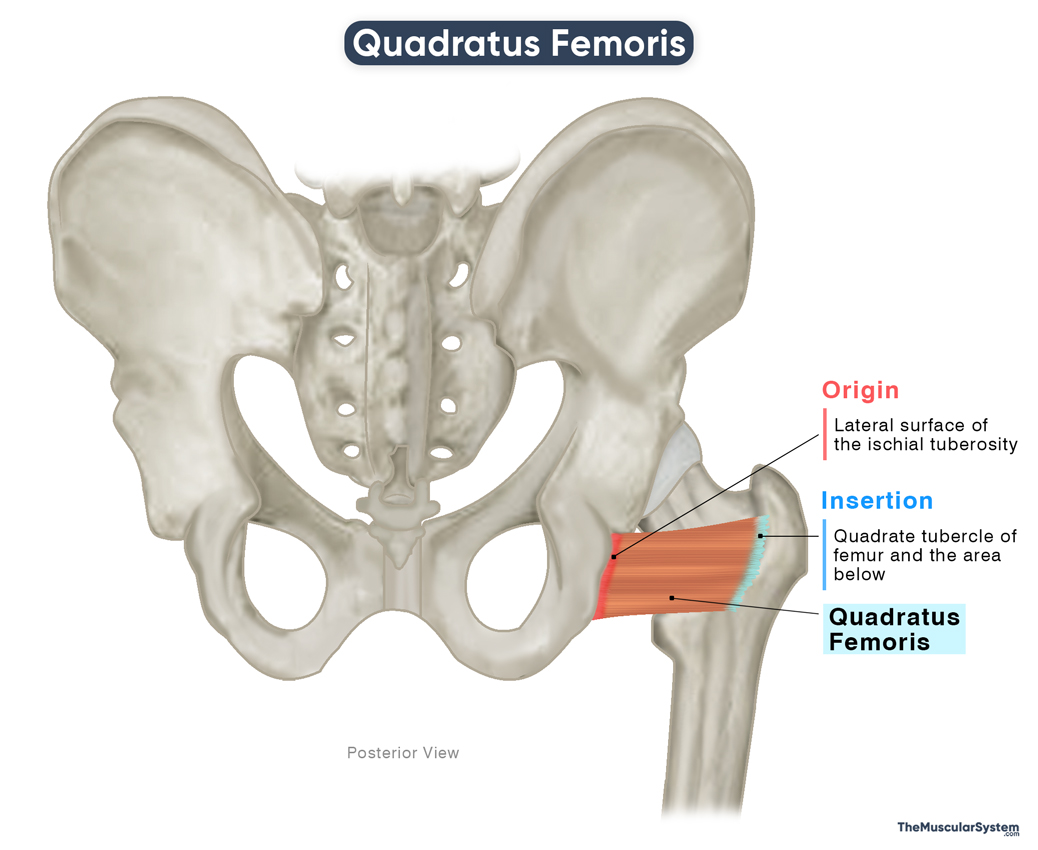
Origin
The quadratus femoris has a broad origin, with its fibers arising from the upper part of the lateral border and the lateral surface of the ischial tuberosity.
Insertion
From the point of origin, the muscle fibers course horizontally and laterally toward the hip joint and the head of the femur (thigh bone). They insert through a broad tendon into the qudrate tubercle of the femur. It is the bony prominence on the intertrochanteric crest, a ridge that connects the greater and lesser tubercles.
Relations With Surrounding Muscles and Structures
The quadratus femoris muscle lies just below the inferior gemellus, with the gluteus maximus covering its posterior surface. Deep and medial to it is the obturator externus, which lies between the hip joint capsule and the quadratus femoris. Inferior and medial to the muscle is the adductor magnus.
The quadrilateral shape of the quadratus femoris gives it nearly horizontal fibers, which run roughly parallel to the fibers of the inferior gemellus above and the adductor magnus below. A gap often exists between the quadratus femoris and the adductor magnus, through which the medial circumflex femoral artery passes.
The sciatic nerve crosses over the posterior aspect of the muscle, particularly near its proximal part. At its distal end, a bursa is commonly present between the muscle and adjacent bony structures, usually near the lesser trochanter, to reduce friction.
Quadratus Femoris Function
| Action | Rotating the thigh laterally at the hip joint |
Rotating the thigh externally: The quadratus femoris is one of the primary external (lateral) rotators of the thigh at the hip joint. It works together with the other deep lateral rotators to rotate the thigh and leg outward. An example of this movement is turning your leg outward to change direction while walking.
Adducting the thigh: Although not its primary function, the quadratus femoris may assist the adductor muscles in bringing the thigh toward the midline (adduction), as in crossing one leg over the other while standing.
Stabilizing the hip joint: Like the other deep gluteal muscles, the quadratus femoris helps stabilize the hip joint by keeping the femoral head securely positioned in the acetabulum through its attachment to the proximal femur.
Antagonists
As one of the major lateral rotators of the thigh, its actions are antagonistic to those of the medial rotators, which include the gluteus medius and minimus, and the tensor fasciae latae muscles.
Innervation
| Nerve | Nerve to quadratus femoris (L4-S1) |
The muscle receives its innervation from the nerve to quadratus femoris, which branches from the anterior divisions of the 4th and 5th lumbar and 1st sacral nerve roots of the sacral plexus. This nerve also supplies the inferior gemellus and sends branches to the hip joint.
Blood Supply
| Artery | Inferior gluteal and medial circumflex femoral arteries |
The muscle has two sources of blood supply. The first is the inferior gluteal artery, which arises from the internal iliac artery and supplies several muscles in the hip and thigh.
Additionally, the muscle receives blood from the descending branch of the medial circumflex femoral artery, a branch of the deep femoral artery, which itself originates from the external iliac artery.
References
- Quadratus Femoris Muscle: Radiopaedia.org
- Quadratus Femoris Muscle: Elsevier.com
- Quadratus Femoris: TeachMeAnatomy.info
- Quadratus Femoris Muscle: Kenhub.com
- Quadratus Femoris – Attachments, Actions & Innervation: GetBodySmart.com
- Quadratus Femoris Muscle: IMAIOS.com
Della Barnes, an MS Anatomy graduate, blends medical research with accessible writing, simplifying complex anatomy for a better understanding and appreciation of human anatomy.
- Latest Posts by Della Barnes, MS Anatomy
-
Tensor Tympani
- -
Stapedius
- -
Auricularis Posterior
- All Posts


