

Longus Colli
By
Della Barnes, an MS Anatomy graduate, blends medical research with accessible writing, simplifying complex anatomy for a better understanding and appreciation of human anatomy.
Last updated:
29/01/2026Della Barnes, MS Anatomy
UX/UI Designer at - AdobeDella Barnes, an MS Anatomy graduate, blends medical research with accessible writing, simplifying complex anatomy for a better understanding and appreciation of human anatomy.
What is the Longus Colli
The longus colli is a long muscle located in the neck, on both sides of the cervical spine. It is one of the four prevertebral muscles of the cervical region, along with the longus capitis, rectus capitis anterior, and rectus capitis lateralis.
The name longus colli comes from Latin, meaning “long muscle of the neck,” with colli translating to “of the neck.” This muscle assists in flexion and rotation of the neck.
Anatomy
Location and Attachments
The muscle runs up the entire length of the cervical spine, from the third thoracic vertebra (T3) to the atlas bone at the top of the cervical spine. It is divided into three parts based on the location and orientation of the muscle fibers.
| Origin | Front of the third cervical to the third thoracic vertebrae (C3-T3) |
| Insertion | Front of the atlas vertebra to the sixth cervical vertebra (C1-C6) |
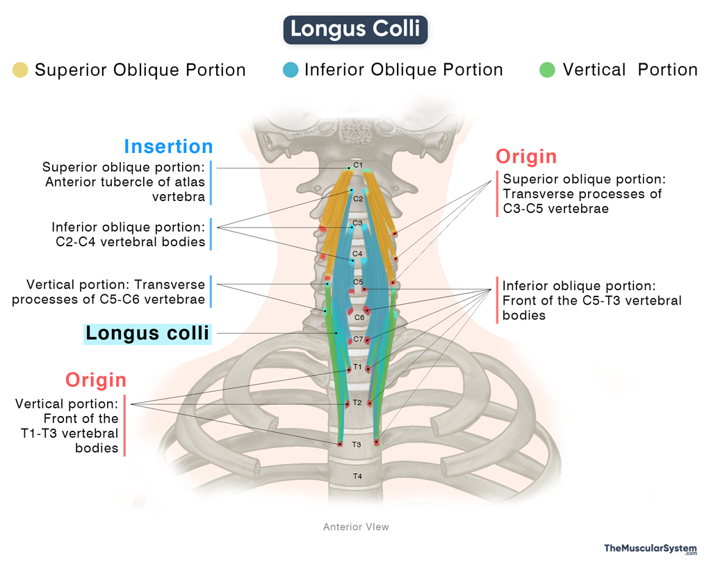
Superior Oblique Portion
It is the superior part of the muscle that originates via narrow tendons from the anterior tubercles of the transverse processes of the third to fifth cervical vertebrae (C3-C5). The fibers ascend obliquely as they form a fleshy belly and finally insert into the anterior tubercle of the atlas vertebra (C1).
Inferior Oblique Portion
It is the middle part of the muscle, originating from the front of the fifth cervical to the third thoracic vertebral bodies (C5-T3). Like the superior portion, it also courses obliquely upward as it forms a belly. As it reaches the higher cervical vertebrae, the belly again becomes tendinous to insert into the second to fourth cervical vertebral bodies (C2-C4).
Vertical Portion
It is the lowest part of the muscle, originating from the front of the first to third thoracic vertebral bodies (T1-T3). Unlike the above two parts, the inferior fibers ascend vertically as they narrow into tendons that insert into the anterior tubercles of the transverse processes of the fifth and sixth cervical vertebrae (C5-C6).
Relations With Surrounding Muscles and Structures
The longus colli, along with the other prevertebral muscles, is surrounded by the prevertebral layer of the deep cervical fascia. All three parts of the muscle are positioned along the groove between the bodies and the transverse processes of the cervical vertebrae. So, in the neck region, the trachea, thyroid gland, esophagus, and all the suprahyoid and infrahyoid muscles lie in front of the longus colli.
The longus colli also contributes to the formation of the scalenovertebral triangle, an opening deep in the neck that serves as a passage for several important neurovascular structures. These include the cervical sympathetic trunk, thoracic duct, and preforaminal part of the vertebral artery (V1). Along its medial aspect course, the subclavian artery, cervical plexus, and brachial plexus.
Function
| Action | Flexing the neck and stabilizing the cervical spine |
It is a weak deep flexor of the neck that assists the stronger deep neck muscles, such as the longus capitis and the scalenes.
Bilateral contraction: When the muscle contracts on both sides, it flexes the neck and bends the head forward, like when you nod your head, bringing the chin toward the chest.
Unilateral contraction: When it contracts on one side, the longus colli helps tilt the head toward the same side (ipsilateral flexion) and rotate it slightly to the opposite side.
Although its small size and nearly sagittal position make it impossible to generate strong movements, the longus colli plays a vital role in stabilizing the neck during motion and in maintaining an upright head posture. The ascending pharyngeal, inferior thyroid, and vertebral arteries supply all three parts of the muscle.
Antagonists
Since the muscle only assists other stronger muscles in their actions, it does not have any direct antagonists.
Innervation
| Nerve | C2-C6 spinal nerves |
Innervation to this muscle comes from the ventral rami of the second to sixth cervical nerves (C2-C6).
Blood Supply
| Artery | Ascending pharyngeal, inferior thyroid, and vertebral arteries |
The ascending pharyngeal, inferior thyroid, and vertebral arteries supply all three parts of the muscle.
References
- Longus Colli Muscle: Radiopaedia.org
- Longus Colli Muscle: Elsevier.com
- Longus Colli Muscle: Kenhub.com
- Longus Colli Muscle Definition, Origin & Insertion: Study.com
- Longus Colli Muscle: IMAIOS.com
Della Barnes, an MS Anatomy graduate, blends medical research with accessible writing, simplifying complex anatomy for a better understanding and appreciation of human anatomy.
- Latest Posts by Della Barnes, MS Anatomy
-
Tensor Tympani
- -
Stapedius
- -
Auricularis Posterior
- All Posts


