

Gluteal Muscles
By
Della Barnes, an MS Anatomy graduate, blends medical research with accessible writing, simplifying complex anatomy for a better understanding and appreciation of human anatomy.
Last updated:
30/10/2025Della Barnes, MS Anatomy
UX/UI Designer at - AdobeDella Barnes, an MS Anatomy graduate, blends medical research with accessible writing, simplifying complex anatomy for a better understanding and appreciation of human anatomy.
What Are the Gluteal Muscles
The gluteal region, or buttock, refers to the area of the body located behind the pelvis and beneath the iliac crest. The muscles located in this region are collectively referred to as the gluteal muscles. In a narrower sense, gluteal muscles, or “glutes,” often refer only to the three large muscles that shape the buttocks: the gluteus maximus, medius, and minimus.
In broader anatomical terms, the gluteal muscles include all the muscles situated in the gluteal region. These muscles help stabilize the pelvis and hips while also playing a crucial role in movements of the hip joint, enabling activities such as walking, running, and more.
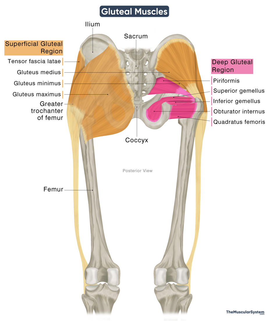
Based on their depth, the gluteal muscles are grouped into two layers: the superficial and the deep gluteal muscles.
Name, Location, & Anatomy of the Gluteal Muscles
Superficial Gluteal Region
It is a group of four muscles that form the superficial layer of the buttocks and is primarily responsible for their shape and contour.
They act primarily to abduct the thigh at the hip joint. Except for the gluteus maximus, the other superficial gluteal muscles also medially rotate the thigh at the hip joint. By attaching to both the ilium and the proximal femur, they also play a crucial role in stabilizing the pelvis and hips, which is essential for maintaining posture and facilitating movement.
Here is a list of the muscles in the superficial gluteal region, with their basic anatomy and functions:
| Muscle | Origin | Insertion | Action | Innervation | Blood Supply |
|---|---|---|---|---|---|
| Gluteus maximus (The largest muscle in the body) | — Gluteal surface of the ilium between the iliac crest and the posterior gluteal line — Posterolateral surfaces of sacrum and coccyx — Thoracolumbar fascia — Sacrotuberous ligament | — Iliotibial tract — Gluteal tuberosity of the femur | — Extending, externally rotating, and abducting (minor) the thigh at the hip joint | Inferior gluteal nerve (L5-S2) | Superior and inferior gluteal arteries |
| Gluteus medius | Gluteal surface of the ilium, between the iliac crest and anterior gluteal line | Greater trochanter of the femur | — Abducting and internally rotating the thigh at the hip joint — Stabilizing the pelvis and hips | Superior gluteal nerve (L4-S1) | Superior gluteal artery |
| Gluteus minimus | Gluteal surface of the ilium, between the anterior and inferior gluteal lines | Greater trochanter of the femur | — Abducting and internally rotating the thigh at the hip joint — Stabilizing the pelvis and hips | Superior gluteal nerve (L4-S1) | Superior gluteal artery |
| Tensor fascia latae | — Anterior superior iliac spine — Outer lip of the iliac crest | Gerdy’s tubercle via the iliotibial tract | — Abducting, medially rotating, and flexing the thigh at the hip joint — Externally rotating the lower leg at the knee joint — Stabilizing the hip and knee | Superior gluteal nerve (L4-S1) | — Lateral circumflex femoral artery — Superior gluteal artery |
Deep Gluteal Region
The deep gluteal region consists of five small and medium-sized muscles situated beneath the gluteus maximus. These muscles primarily act as lateral rotators of the thigh at the hip joint, but they also contribute to stabilizing the head of the femur within the acetabulum during movement.
Here is a list of the muscles in the deep gluteal region, with their location, functions, and anatomy:
| Muscle | Origin | Insertion | Action | Innervation | Blood Supply |
|---|---|---|---|---|---|
| Piriformis | Anterior surface of sacrum (S2-S4) | Greater trochanter of the femur | — Laterally rotating the thigh at the hip joint — Stabilizing the hip joint — Abducting the thigh when the hip is flexed | Nerve to Piriformis (S1-S2) | — Inferior and superior gluteal arteries — Internal pudendal artery — Lateral sacral artery |
| Obturator internus | — Ischiopubic ramus — Posterior surface of the obturator membrane | Trochanteric fossa of the femur | — Laterally rotating the thigh at the hip joint — Stabilizing the hip joint — Abducting the thigh when the hip is flexed | Nerve to obturator internus (L5-S2) | Inferior gluteal artery |
| Superior gemellus | Posterior surface of the ischial spine | Trochanteric fossa via the obturator internus tendon | Helping the obturator internus in its actions | Nerve to obturator internus (L5-S2) | Inferior gluteal artery |
| Inferior gemellus | Superior-posterior surface of the ischial tuberosity | Trochanteric fossa via the obturator internus tendon | Helping the obturator internus in its actions | Nerve to quadratus femoris (L5-S1) | Inferior gluteal artery |
| Quadratus femoris | Lateral surface of the ischial tuberosity | Quadrate tubercle of the femur | Laterally rotating the thigh at the hip joint | Nerve to quadratus femoris (L4-S1) | — Inferior gluteal artery — Medial circumflex femoral artery |
Note: Although the obturator externus also acts as a lateral rotator of the thigh, it is anatomically part of the medial compartment of the thigh and is therefore not included in the list of gluteal region muscles above.
All the muscles in the gluteal region receive innervation from branches of the sacral plexus, and their blood supply comes primarily from branches of the internal iliac artery. An exception is the quadratus femoris, which also receives blood from the medial circumflex femoral artery, a branch of the deep femoral artery.
Several important nerves and blood vessels pass through the greater sciatic foramen to enter the gluteal region. These include the sciatic nerve, superior and inferior gluteal nerves and vessels, and the posterior cutaneous nerve of the thigh, among others. The piriformis muscle serves as a landmark, with some structures emerging above it, while others pass below.
References
- Muscles of the Gluteal Region: TeachMeAnatomy.info
- Gluteal Muscles (Glutes): Clevelandclinic.org
- Gluteal Region: IMAIOS.com
- Muscles of Gluteal Region: Elsevier.com
- Gluteal Muscles: Radiopaedia.org
Della Barnes, an MS Anatomy graduate, blends medical research with accessible writing, simplifying complex anatomy for a better understanding and appreciation of human anatomy.
- Latest Posts by Della Barnes, MS Anatomy
-
Tensor Tympani
- -
Stapedius
- -
Auricularis Posterior
- All Posts


