

External Anal Sphincter
By
Della Barnes, an MS Anatomy graduate, blends medical research with accessible writing, simplifying complex anatomy for a better understanding and appreciation of human anatomy.
Last updated:
26/06/2025Della Barnes, MS Anatomy
UX/UI Designer at - AdobeDella Barnes, an MS Anatomy graduate, blends medical research with accessible writing, simplifying complex anatomy for a better understanding and appreciation of human anatomy.
What is the External Anal Sphincter
The external anal sphincter is a ring of skeletal muscle that surrounds the lower end of the anal canal. Along with the internal anal sphincter, it forms the sphincter ani muscle complex responsible for controlling defecation. The muscle also belongs to the anal triangle of the perineum.
Since it is composed of skeletal muscle, it is under voluntary control via the somatic nervous system, helping with controlling the opening and closing of the anal canal. In contrast, the internal anal sphincter is made of smooth muscle and functions involuntarily under the autonomic nervous system.
Anatomy
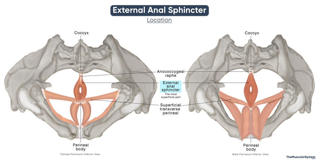
Location and Attachments
It is a layered circular muscle that cannot be described with general muscle anatomy terminology, like “origin” and “insertion.” The muscle surrounds the lower two-thirds of the anal canal, attaching to fascia, ligament, and skin tissue instead of having any significant bony attachments.
Classically, the external anal sphincter is divided into three parts: deep, superficial, and subcutaneous. However, modern anatomical studies have shown that these layers often blend together and are not always clearly distinguishable. Despite this, the three divisions remain a standard approach in basic anatomical education.
1. Deep Part
It is the most proximal or superior part of the external anal sphincter, encircling the highest portion of the anal canal. The circular muscle fibers in this part blend with the puborectalis muscle, the most frontal part of the levator ani, at the back and sides. Some fibers may cross over to the other side of the midline to fuse with the superficial transverse perineal muscle.
The deep part of the external anal sphincter inserts at the back into the anococcygeal raphe, the fibrous band connecting the anus with the coccyx.
2. Superficial Part
It is the middle part of the muscle, between the deep and subcutaneous parts. The fibers attach to the perineal body at the front, and to the coccyx via the anococcygeal raphe at the back. This posterior attachment is the only bony attachment of the external anal sphincter. Some muscle fibers may also cross the midline to attach to the other side.
Unlike the deep part, the fibers in the superficial part are more elliptical than circular in a cross-section.
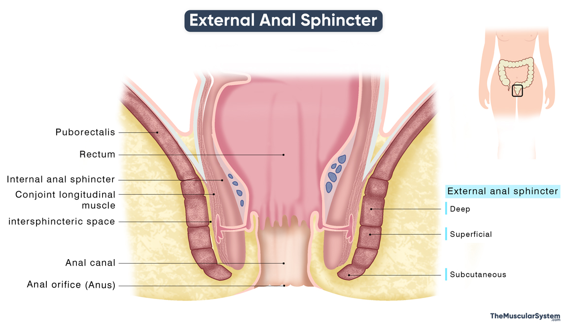
3. Subcutaneous Part
This is the most superficial part of the external anal sphincter, located just beneath the skin. Its muscle fibers are arranged in a circular pattern and encircle the anal verge, the lowest and most external portion of the anal canal.
External Anal Sphincter Relations With Surrounding Muscles and Structures
The muscle lies anterior to the anococcygeal raphe, while the perineal body and the two transverse perineal muscles lie in front of it. Superiorly, it is related to the pelvic floor muscle, the levator ani. The relationships with these surrounding muscles and structures have already been discussed. At the point where the external anal sphincter merges with the puborectalis muscle, they form a muscular ring known as the anorectal ring, which marks the junction between the rectum and the anal canal.
Within the anal triangle, the external anal sphincter, along with the levator ani, forms the medial boundary of the ischioanal fossa, a fat-filled space on either side of the anal canal that allows for expansion during defecation.
The deep and superficial parts of the external anal sphincter overlap with the lowest part of the internal anal sphincter, a ring of smooth muscle that also surrounds the anal canal. In this overlapping region, a smooth muscle called the conjoint longitudinal muscle lies between the two anal sphincters and serves to separate them. Its fibers pass through the subcutaneous and deep parts of the external anal sphincter.
A small space between these two muscles, known as the intersphincteric space, contains the anal glands and serves as an important anatomical and surgical landmark.
Function
| Action | Providing voluntary control over the anal canal |
Role in Anal Opening and Closure
The external and internal anal sphincters work together to control the anal canal and support the process of defecation. Under normal conditions, the external anal sphincter maintains a state of continuous tonic contraction, keeping the anal canal closed and preventing involuntary stool passage.
When the rectum fills with feces, this sphincter can be voluntarily contracted to maintain continence. At an appropriate time and place, it can then be voluntarily relaxed to allow for the anal orifice (anus) to open for defecation. The puborectalis muscle, which forms a sling around the anorectal junction, works with the external sphincter to help keep the anal canal closed.
Role in Pelvic Floor Stability
It works together with the other perineal muscles to help stabilize the pelvic floor.
Antagonists
The external anal sphincter does not have an antagonistic muscle acting against it.
Innervation
| Nerve | Perineal and inferior rectal branches of the pudendal nerve (S2-S4) |
The somatic innervation to the muscle comes from the perineal and inferior rectal (or inferior anal) branches of the pudendal nerve, which arises from the ventral rami of the 2nd to 4th sacral spinal nerves (S2 to S4).
Blood Supply
| Artery | Inferior rectal arteries |
The muscle receives its primary blood supply from the inferior rectal (hemorrhoidal) arteries, which are the terminal branches of the internal pudendal arteries. The internal pudendal arteries, in turn, arise from the internal iliac artery.
References
- Anal Sphincter: Radiopaedia.org
- External Anal Sphincter: Kenhub.com
- External Anal Sphincter: Elsevier.com
- External Anal Sphincter: IMAIOS.com
- Anatomy, Abdomen and Pelvis: Anal Canal: NCBI.NLM.NIH.gov
- The Anal Canal: TeachMeAnatomy.info
Della Barnes, an MS Anatomy graduate, blends medical research with accessible writing, simplifying complex anatomy for a better understanding and appreciation of human anatomy.
- Latest Posts by Della Barnes, MS Anatomy
-
Tensor Tympani
- -
Stapedius
- -
Auricularis Posterior
- All Posts


