

Iliopsoas
By
Della Barnes, an MS Anatomy graduate, blends medical research with accessible writing, simplifying complex anatomy for a better understanding and appreciation of human anatomy.
Last updated:
14/05/2025Della Barnes, MS Anatomy
UX/UI Designer at - AdobeDella Barnes, an MS Anatomy graduate, blends medical research with accessible writing, simplifying complex anatomy for a better understanding and appreciation of human anatomy.
What is the Iliopsoas
The iliopsoas (pronounced ILL-ee-oh-SOH-us) is a composite muscle and the strongest flexor of the hip in humans. It is comprised of two of the largest muscles in the posterior abdominal wall, the psoas major and the iliacus. The name ‘iliopsoas’ is derived from the names of these contributing muscles.
Structure, Anatomy, and Location
The psoas major and iliacus course as separate muscles in the posterior abdomen. Here are the anatomical details of the two muscles:
| Muscle | Origin | Insertion | Innervation | Blood Supply |
| Psoas Major | T12 to L4 vertebrae | Lesser trochanter of the femur | Anterior rami of L1-L3 nerves | Lumbar branch of the iliolumbar artery |
| Iliacus | Iliac crest and upper surface of iliac fossa | Lesser trochanter of the femur | Femoral nerve | Lumbar branch of the iliolumbar artery |
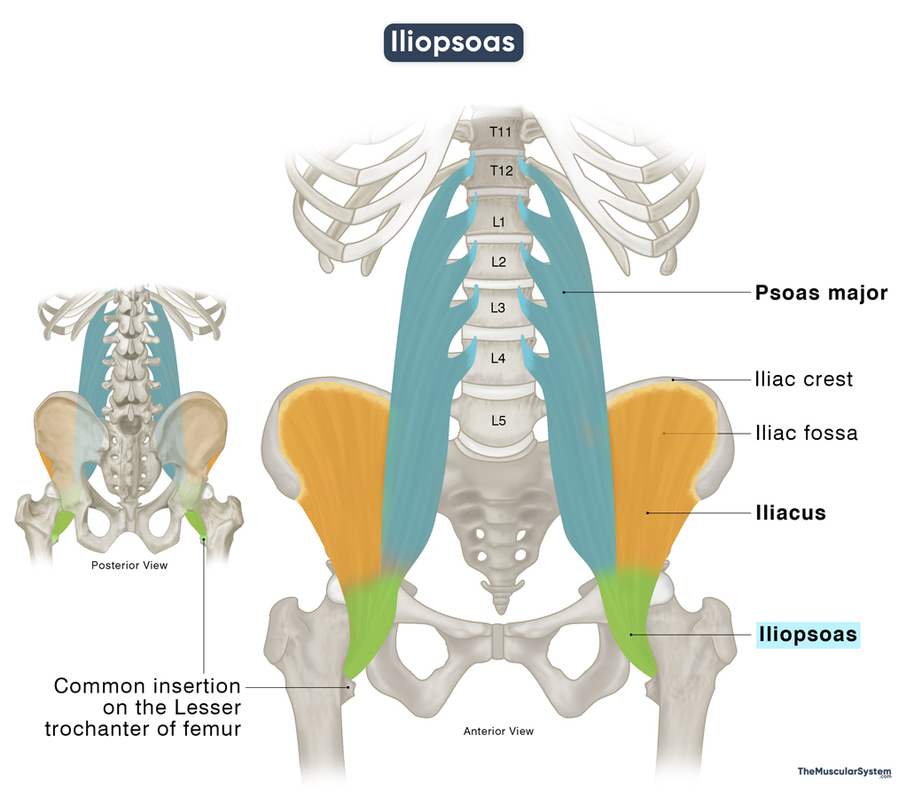
Once these two muscles get to the level of the inguinal ligament in the groin region, they merge to form the iliopsoas muscle. From there, it courses downward to insert via a single thick tendon into the lesser trochanter, the small bony prominence below the neck of the back of the femur bone.
The psoas minor is a muscle of the posterior abdominal wall that is occasionally considered part of the iliopsoas. However, it is present in only about 40-60% of the population. Even when present, it inserts into the iliopubic eminence and does not merge with the iliacus or psoas major. For this reason, it is not considered part of the iliopsoas in this article.
Function
The iliopsoas is essential for the flexibility, movement, and postural stability of the hip, thigh, and lower back. Here are its primary actions:
1. Flexing the Hip and Thigh
The iliopsoas is the strongest hip flexor. Both the psoas major and the iliacus are powerful flexors of the thigh and hip. When they contract together as the iliopsoas, they produce even stronger hip flexion, bringing the thigh closer to the torso. This movement occurs during actions such as high-knee running or kicking.
2. Maintaining Posture
When standing upright, the iliopsoas muscle helps maintain normal lumbar lordosis, the natural inward curve of the lower back. It exerts a slight forward and downward pull on the lumbar spine, contributing to the preservation of this spinal alignment.
By maintaining lumbar lordosis, the iliopsoas indirectly supports the compensatory thoracic kyphosis (the outward curve of the thoracic spine in the mid back), preserving the spine’s natural curvature.
Antagonists
The primary antagonist of the iliopsoas is the gluteus maximus, the largest muscle in the human body. While the iliopsoas flexes the hip, the gluteus maximus extends it, making them functionally opposing muscles during movements such as walking, running, or climbing stairs.
References:
- Iliopsoas: TeachMeAnatomy.info
- Iliopsoas Muscle: Kenhub.com
- Anatomy, Bony Pelvis and Lower Limb, Iliopsoas Muscle: NCBI.NLM.NIH.gov
- Iliopsoas Muscle: Radiopaedia.org
- Iliopsoas Muscle: GetBodySmart.com
Della Barnes, an MS Anatomy graduate, blends medical research with accessible writing, simplifying complex anatomy for a better understanding and appreciation of human anatomy.
- Latest Posts by Della Barnes, MS Anatomy
-
Tensor Tympani
- -
Stapedius
- -
Auricularis Posterior
- All Posts


